Pineocytoma MRI: Difference between revisions
Homa Najafi (talk | contribs) (Created page with "__NOTOC__ {{Pineocytoma}} {{CMG}}{{AE}}{{SR}}{{Homa}} ==Overview== There are no MRI findings associated with [disease name]. OR [Location] MRI may be helpful in the diagnos...") |
Homa Najafi (talk | contribs) No edit summary |
||
| Line 31: | Line 31: | ||
*[Complication 2] | *[Complication 2] | ||
*[Complication 3] | *[Complication 3] | ||
===MRI=== | |||
*Brain MRI may be diagnostic of pineocytoma. | |||
*Features on MRI suggestive of pineocytoma include:<ref name="scan1">Radiographic features of pineocytoma. Dr Bruno Di Muzio and Dr Frank Gaillard et al. Radiopeadia 2015. http://radiopaedia.org/articles/pineocytoma. Accessed on November 20, 2015</ref> | |||
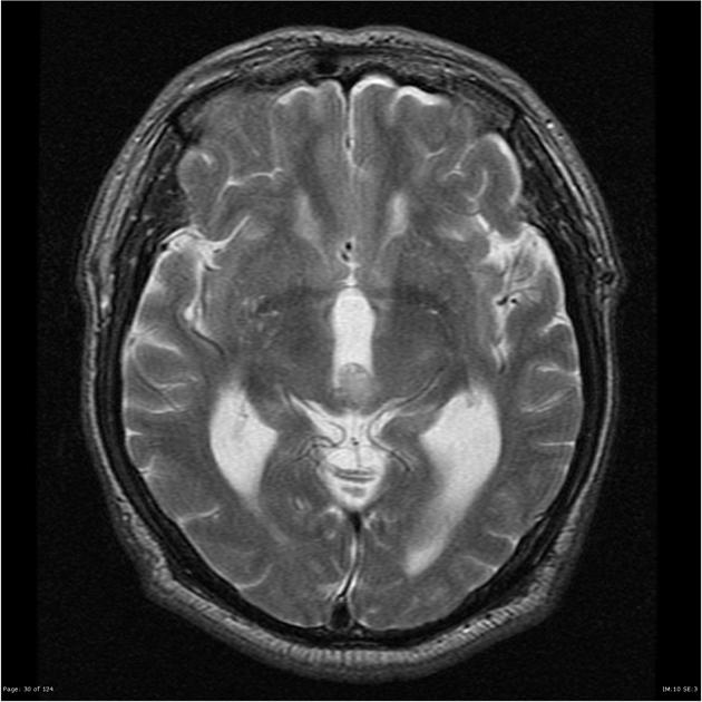
[[File:MRI image of pineocytoma 2.jpg|center|thumb|A large and ill-defined mass is present in the region of the pineal gland, demonstrating contrast enhancement.<ref name="mri1">Image courtesy of Dr. Frank Gaillard. Radiopaedia (original file [http://radiopaedia.org/cases/pineocytoma-1 here]). Creative Commons BY-SA-NC</ref>]] | |||
{| style="border: 0px; font-size: 90%; margin: 3px; width:1000px" | |||
| valign="top" | | |||
|+ | |||
! style="background: #4479BA; width: 300px;" | {{fontcolor|#FFF|MRI component}} | |||
! style="background: #4479BA; width: 700px;" | {{fontcolor|#FFF|Findings}} | |||
|- | |||
| style="padding: 5px 5px; background: #DCDCDC; font-weight: bold" align="center" | | |||
T1 | |||
| style="padding: 5px 5px; background: #F5F5F5;" | | |||
*Isointense to brain parenchyma | |||
|- | |||
| style="padding: 5px 5px; background: #DCDCDC;font-weight: bold" align="center" | | |||
T2 | |||
| style="padding: 5px 5px; background: #F5F5F5;" | | |||
*Solid components are isointense to brain parenchyma | |||
*Areas of cystic change | |||
*Sometimes the majority of the tumor is cystic | |||
|- | |||
| style="padding: 5px 5px; background: #DCDCDC;font-weight: bold" align="center" | | |||
T1 with gadolinium contrast | |||
| style="padding: 5px 5px; background: #F5F5F5;" | | |||
*Solid components vividly enhance | |||
|} | |||
==References== | ==References== | ||
Latest revision as of 16:02, 10 October 2019
Editor-In-Chief: C. Michael Gibson, M.S., M.D. [1]Associate Editor(s)-in-Chief: Sujit Routray, M.D. [2] Homa Najafi, M.D.[3]
Overview
There are no MRI findings associated with [disease name].
OR
[Location] MRI may be helpful in the diagnosis of [disease name]. Findings on MRI suggestive of/diagnostic of [disease name] include [finding 1], [finding 2], and [finding 3].
OR
There are no MRI findings associated with [disease name]. However, a MRI may be helpful in the diagnosis of complications of [disease name], which include [complication 1], [complication 2], and [complication 3].
MRI
There are no MRI findings associated with [disease name].
OR
[Location] MRI may be helpful in the diagnosis of [disease name]. Findings on MRI suggestive of/diagnostic of [disease name] include:
- [Finding 1]
- [Finding 2]
- [Finding 3]
OR
There are no MRI findings associated with [disease name]. However, a MRI may be helpful in the diagnosis of complications of [disease name], which include:
- [Complication 1]
- [Complication 2]
- [Complication 3]
MRI
- Brain MRI may be diagnostic of pineocytoma.
- Features on MRI suggestive of pineocytoma include:[1]

| MRI component | Findings |
|---|---|
|
T1 |
|
|
T2 |
|
|
T1 with gadolinium contrast |
|
References
- ↑ Radiographic features of pineocytoma. Dr Bruno Di Muzio and Dr Frank Gaillard et al. Radiopeadia 2015. http://radiopaedia.org/articles/pineocytoma. Accessed on November 20, 2015
- ↑ Image courtesy of Dr. Frank Gaillard. Radiopaedia (original file here). Creative Commons BY-SA-NC