Patent ductus arteriosus pathophysiology: Difference between revisions
m (Robot: Automated text replacement (-msbeih@perfuse.org +msbeih@wikidoc.org, -psingh@perfuse.org +psingh13579@gmail.com, -agovi@perfuse.org +agovi@wikidoc.org, -rgudetti@perfuse.org +ravitheja.g@gmail.com, -lbiller@perfuse.org +lbiller@wikidoc.org,...) |
|||
| Line 24: | Line 24: | ||
##A decrease in the relative size of the ductus compared with other cardiovascular structures. This results in a medium-sized defect compared with the course expected for a medium-sized defect. | ##A decrease in the relative size of the ductus compared with other cardiovascular structures. This results in a medium-sized defect compared with the course expected for a medium-sized defect. | ||
##The development of severe pulmonary vascular obstructive disease, can occur at any time from age 3 until early adulthood. The [[left-to-right shunt]] converts to a [[right-to-left shunt]] with [[cyanosis]] and disappearance of the [[continuous murmur]]. | ##The development of severe pulmonary vascular obstructive disease, can occur at any time from age 3 until early adulthood. The [[left-to-right shunt]] converts to a [[right-to-left shunt]] with [[cyanosis]] and disappearance of the [[continuous murmur]]. | ||
Shown below is the pictoral image of pathophysiology of patent ductus arteriosus | Shown below is the pictoral image of pathophysiology of patent ductus arteriosus | ||
[[Image:Pathophysiology_of_patent_ductus_arteriosus_2.jpg|center|500px|Pictoral illustration of patent ductus arteriosus]] | [[Image:Pathophysiology_of_patent_ductus_arteriosus_2.jpg|center|500px|Pictoral illustration of patent ductus arteriosus]] | ||
----- | |||
Shown below is the image of pathophysiology of patent ductus arteriosus in the cross-section of the heart | Shown below is the image of pathophysiology of patent ductus arteriosus in the cross-section of the heart | ||
[[Image:Pathophysiology_of_patent_ductus_arteriosus.jpg|center|500px|Pathophysiology of PDA]] | [[Image:Pathophysiology_of_patent_ductus_arteriosus.jpg|center|500px|Pathophysiology of PDA]] | ||
---- | |||
===Gross Pathology=== | |||
<div align="left"> | |||
<gallery heights="175" widths="175"> | |||
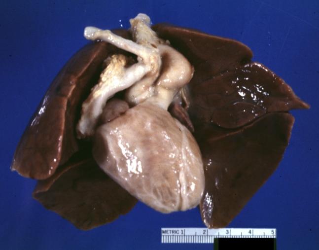
Image:821.jpg|Patent Ductus Arteriosus: Gross example in an infant heart | |||
Image:4433.jpg|Patent Ductus Arteriosus: Gross fixed tissue probe in ductus | |||
</gallery> | |||
</div> | |||
<div align="left"> | |||
<gallery heights="175" widths="175"> | |||
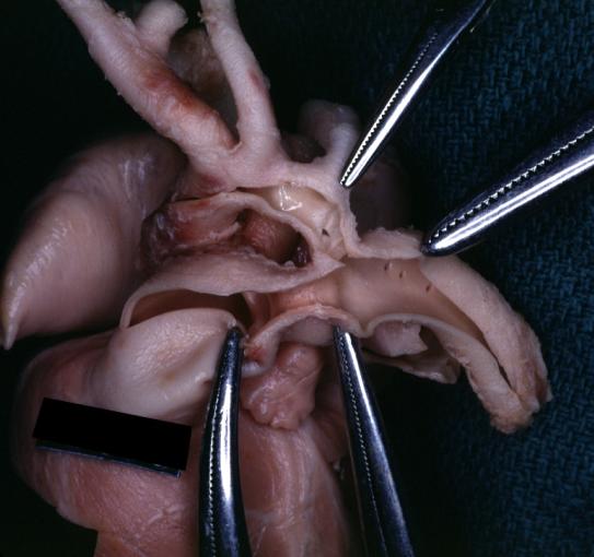
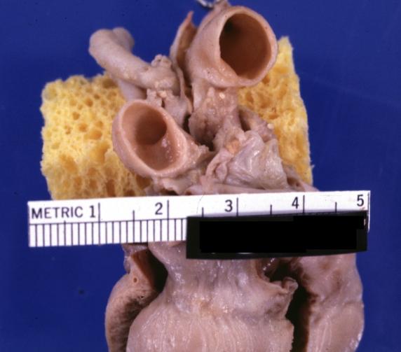
Image:5320.jpg|Patent Ductus Arteriosus: Gross fixed tissue view of ductus opened from pulmonary artery into aorta with edematous appearing intimal surface | |||
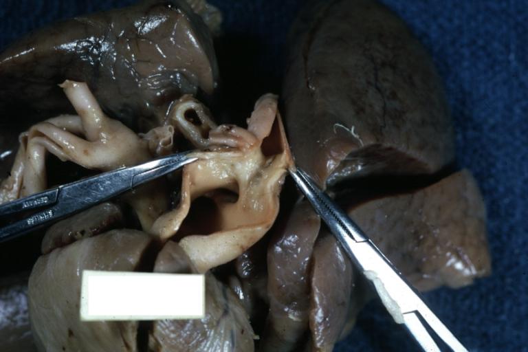
Image:5338.jpg|Patent Ductus Arteriosus: Gross natural color opened ductus in infant shows apparent intimal edema in ductus. | |||
</gallery> | |||
</div> | |||
<div align="left"> | |||
<gallery heights="175" widths="175"> | |||
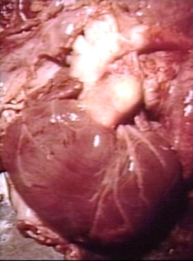
Image:6818.jpg|Patent Ductus Arteriosus with Aneurysmal Dilation: Gross fixed tissue external photo of heart shows the lesion | |||
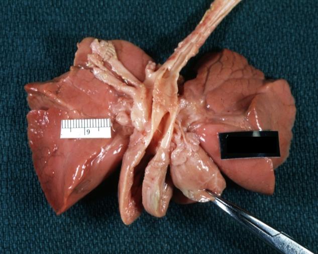
Image:6819.jpg|Patent Ductus Arteriosus with Aneurysmal Dilation: Gross fixed tissue aorta and ductus have been cross sectioned showing arch of aorta and huge ductus in a 5 day old infant | |||
</gallery> | |||
</div> | |||
<div align="left"> | |||
<gallery heights="175" widths="175"> | |||
Image:6820.jpg|Patent Ductus Arteriosus with Aneurysmal Dilation: Gross fixed tissue opened aortic arch and descending thoracic showing very large opening of ductus into aorta | |||
Image:249019.jpg|Patent Ductus Arteriosus | |||
</gallery> | |||
</div> | |||
===Videos=== | |||
{{#ev:youtube|5_BNHYGUgG4}} | {{#ev:youtube|5_BNHYGUgG4}} | ||
Revision as of 15:28, 3 January 2013
|
Patent Ductus Arteriosus Microchapters |
|
Differentiating Patent Ductus Arteriosus from other Diseases |
|---|
|
Diagnosis |
|
Treatment |
|
Medical Therapy |
|
Case Studies |
|
Patent ductus arteriosus pathophysiology On the Web |
|
American Roentgen Ray Society Images of Patent ductus arteriosus pathophysiology |
|
Risk calculators and risk factors for Patent ductus arteriosus pathophysiology |
Editor-In-Chief: C. Michael Gibson, M.S., M.D. [1]; Associate Editor-In-Chief: Priyamvada Singh, M.B.B.S. [2], Cafer Zorkun, M.D., Ph.D. [3], Assistant Editor-In-Chief: Kristin Feeney, B.S. [4]
Overview
The pathophysiological findings depend on the size of defect and the pulmonary vascular resistance
Pathophysiology
The pathophysiological consequences depend on the size of the defect and the pulmonary vascular resistance. [1]
Small PDA
- Small left-to-right shunt (Qp/Qs < 1.5).
- Normal ratio of pulmonary artery (PA) to systemic pressure.
- Shunt throughout the cardiac cycle, continuous murmur.
Medium-sized PDA
- Qp/Qs 1.5 to 2.0 yet small enough to offer some resistance to flow.
- PA systolic to systemic pressures are < 0.5.
- Unusual for this group to have markedly increased PVR.
- Due to increased return to the left heart, there is volume overload of the left atrium (LA) and the left ventricle (LV).
Large PDA
- Defect does not restrict flow.
- There is pulmonary hypertension at near systemic pressures (PA systolic/systolic pressure is >0.5).
- Because of the physiologic decrease in the PVR over the first three months of life there is a large left-to-right shunt with Qp/Qs > 2.
- The large volume overload of the left ventricle may result in LV failure.
- There is pulmonary hypertension.
- There may be two courses:
- A decrease in the relative size of the ductus compared with other cardiovascular structures. This results in a medium-sized defect compared with the course expected for a medium-sized defect.
- The development of severe pulmonary vascular obstructive disease, can occur at any time from age 3 until early adulthood. The left-to-right shunt converts to a right-to-left shunt with cyanosis and disappearance of the continuous murmur.
Shown below is the pictoral image of pathophysiology of patent ductus arteriosus

Shown below is the image of pathophysiology of patent ductus arteriosus in the cross-section of the heart

Gross Pathology
-
Patent Ductus Arteriosus: Gross example in an infant heart
-
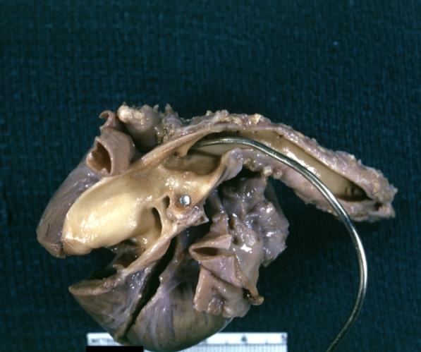
Patent Ductus Arteriosus: Gross fixed tissue probe in ductus


-
Patent Ductus Arteriosus: Gross fixed tissue view of ductus opened from pulmonary artery into aorta with edematous appearing intimal surface
-
Patent Ductus Arteriosus: Gross natural color opened ductus in infant shows apparent intimal edema in ductus.


-
Patent Ductus Arteriosus with Aneurysmal Dilation: Gross fixed tissue external photo of heart shows the lesion
-
Patent Ductus Arteriosus with Aneurysmal Dilation: Gross fixed tissue aorta and ductus have been cross sectioned showing arch of aorta and huge ductus in a 5 day old infant


-
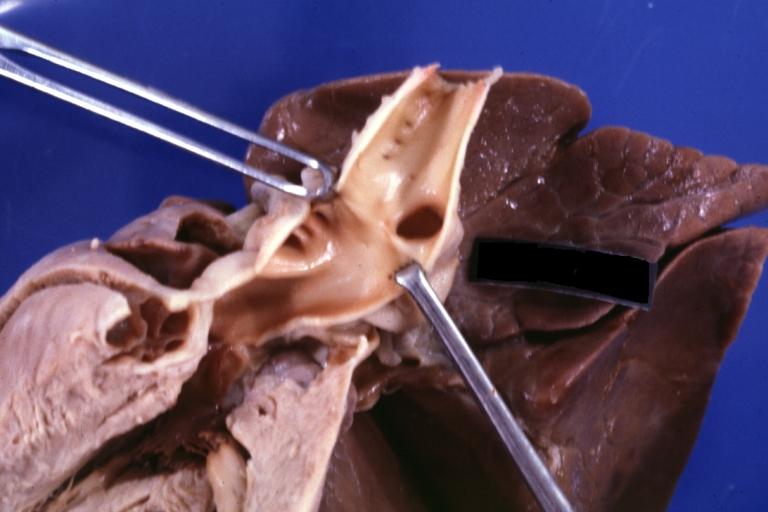
Patent Ductus Arteriosus with Aneurysmal Dilation: Gross fixed tissue opened aortic arch and descending thoracic showing very large opening of ductus into aorta
-
Patent Ductus Arteriosus


Videos
{{#ev:youtube|5_BNHYGUgG4}}
References
- ↑ Giuliani et al, Cardiology: Fundamentals and Practice, Second Edition, Mosby Year Book, Boston, 1991, pp. 1653-1663.