Parkes Weber syndrome
Editor-In-Chief: C. Michael Gibson, M.S., M.D. [1]
Associate Editor-In-Chief: Cafer Zorkun, M.D., Ph.D. [2]
Overview
Parkes Weber Syndrome also known as (PKWS) is a rare vascular abnormality characterized by a cutaneous flush with underlying multiple CM (capillary malformation), VM (venuous malformation), LM (lymphatic malformation) and AVFs (arteriovenous fistulas), in association with soft tissue and skeletal hypertrophy of the affected limb. [1] [2][3][4] This disease is clinically distinct from Klippel-Trenaunay syndrome but the two are often confused as the presentation is very similar. AVMs present in PWS serve as the hallmark for distinguishing the two syndromes.[3][5]
Discovered by Frederick P. Weber in 1907, who noted two patients with port-wine stains and enlargement of the limb with accompanying enlargement of the vasculature leading to a palpable thrill. Recently a disorder in the RASA1 gene has been implicated in development of this syndrome.[6] F. Parkes Weber's (1863-1962) name is also attached to hereditary hemorrhagic telangiectasia, Sturge-Weber syndrome, Weber-Christian disease, and Klippel-Trenaunay-Weber syndrome.
Historical Perspective
- Parkes-Weber Syndrome was first discovered by Frederick Parkes Weber, an English dermatologist, in 1907.
- In 2003, RASA1 mutations were first identified in the pathogenesis of this syndrome.
- In [year], the first [discovery] was developed by [scientist] to treat/diagnose [disease name].
Classification
- [Disease name] may be classified according to [classification method] into [number] subtypes/groups:
- [group1]
- [group2]
- [group3]
- Other variants of [disease name] include [disease subtype 1], [disease subtype 2], and [disease subtype 3].
Pathophysiology
Gene map locus is 5q13.3.
Six families reported by Eerola et al. in 2003, manifested atypical capillary malformations associated with either arteriovenous malformation, arteriovenous fistula, or Parkes Weber syndrome. They named this association CM-AVM for 'capillary malformation-arteriovenous malformation' and found mutation in the RASA1 gene in affected members of these families.
- The pathogenesis of [disease name] is characterized by [feature1], [feature2], and [feature3].
- The [gene name] gene/Mutation in [gene name] has been associated with the development of [disease name], involving the [molecular pathway] pathway.
- On gross pathology, [feature1], [feature2], and [feature3] are characteristic findings of [disease name].
- On microscopic histopathological analysis, [feature1], [feature2], and [feature3] are characteristic findings of [disease name].
Clinical Features
PWS can present with:
- Upper or lower limb hypertrophy
- Abnormal bleeding/ recurrent bleeding from skin lesions
- Large, flat, pink discolouration on the skin known as a port-wine stain (naevus flammeus)
- Enlarged arteries and veins showing evidence of high flow AV shunting.
- Capillary malformations resulting in petecheia on face, arms and legs
- Telangiectasia
- AV fistulas
- Venous malformations
- Varicose veins
- Congestive heart failure
Differentiating Parkes-Weber Syndrome from other Diseases
- Parkes Weber Syndrome must be differentiated from other diseases that cause port-wine stains and capillary malformations, such as:
- Klippel-Trenaunay Syndrome
- Sturge-Weber syndrome: Sturge-Weber syndrome is characterized by a triad of facial port wine stain (classically in the distribution area of the ophthalmic and/or maxillary branch (segments V1/V2) of the trigeminal nerve), leptomeningeal angiomatosis, and ocular involvement. The capillary malformation associated with SWS usually presents as both upper and lower eyelid staining, and is often bilateral.[7]
- Proteus syndrome — Proteus syndrome is an extremely rare disorder characterized by random overgrowth of body parts. The cause is considered to be mosaicism for a somatic activating mutation in the AKT1 oncogene.
- CLOVES syndrome
- Capillary malformation-arteriovenous malformation syndrome
Epidemiology and Demographics
- Exact prevalence of PWS is unknown.[4]
Natural History, Complications and Prognosis
- Early clinical features include
- If left untreated, [#%] of patients with [disease name] may progress to develop [manifestation 1], [manifestation 2], and [manifestation 3].
- Common complications of [disease name] include [complication 1], [complication 2], and [complication 3].
- Prognosis is generally [excellent/good/poor], and the [1/5/10year mortality/survival rate] of patients with [disease name] is approximately [#%].
Diagnosis
Diagnostic Criteria
- The diagnosis of [disease name] is made when at least [number] of the following [number] diagnostic criteria are met:
- [criterion 1]
- [criterion 2]
- [criterion 3]
- [criterion 4]
Symptoms
- [Disease name] is usually asymptomatic.
- Symptoms of [disease name] may include the following:
- [symptom 1]
- [symptom 2]
- [symptom 3]
- [symptom 4]
- [symptom 5]
- [symptom 6]
Physical Examination
- Patients with [disease name] usually appear [general appearance].
- Physical examination may be remarkable for:
- [finding 1]
- [finding 2]
- [finding 3]
- [finding 4]
- [finding 5]
- [finding 6]
Laboratory Findings
- There are no specific laboratory findings associated with [disease name].
- A [positive/negative] [test name] is diagnostic of [disease name].
- An [elevated/reduced] concentration of [serum/blood/urinary/CSF/other] [lab test] is diagnostic of [disease name].
- Other laboratory findings consistent with the diagnosis of [disease name] include [abnormal test 1], [abnormal test 2], and [abnormal test 3].
Imaging Findings
- There are no [imaging study] findings associated with [disease name].
- [Imaging study 1] is the imaging modality of choice for [disease name].
- On [imaging study 1], [disease name] is characterized by [finding 1], [finding 2], and [finding 3].
- [Imaging study 2] may demonstrate [finding 1], [finding 2], and [finding 3].
Other Diagnostic Studies
- [Disease name] may also be diagnosed using [diagnostic study name].
- Findings on [diagnostic study name] include [finding 1], [finding 2], and [finding 3].
Treatment
Medical Therapy
- There is no treatment for PWS; the mainstay of therapy is supportive care.
Surgery
- Surgery is the mainstay of therapy for [disease name].
- [Surgical procedure] in conjunction with [chemotherapy/radiation] is the most common approach to the treatment of [disease name].
- [Surgical procedure] can only be performed for patients with [disease stage] [disease name].
Prevention
- PWS is a genetic disease as such there are no primary preventive measures available.
- Once diagnosed and successfully treated, patients with [disease name] are followed-up every [duration]. Follow-up testing includes [test 1], [test 2], and [test 3].
Multi Sliced CT
-
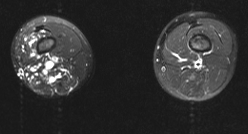
Multi Sliced CT - Lower extremities: A 10 year old boy presented for assessment of leg length discrepancy and cutaneous “capillary” vascular malformation. The provisional diagnosis was Klippel-Trenaunay syndrome. The axial fat-saturated T2 weighted MRI above shows dilated vascular structures in the right lower limb involving subcutaneous and multiple muscle compartments. Flow voids and pulsation artefact (particularly laterally) suggest a high flow component consistent with arteriovenous malformation. These findings favour Parkes Weber syndrome over Klippel-Trenaunay syndrome. There was no high output cardiac failure in this case. (Image courtesy of Dr Laughlin Dawes)

See Also
References
- ↑ Eerola, I.; Boon, L. M.; Mulliken, J. B.; Burrows, P. E.; Dompmartin, A.; Watanabe, S.; Vanwijck, R.; Vikkula, M. Capillary malformation-arteriovenous malformation, a new clinical and genetic disorder caused by RASA1 mutations. Am. J. Hum. Genet. 73: 1240-1249, 2003. PMID 14639529
- ↑ Mulliken, J. B.; Young, A. E. (eds.): Vascular Birthmarks: Hemangiomas and Vascular Malformations. Philadelphia: W. B. Saunders Co., 1988
- ↑ 3.0 3.1 Lee BB. (2012). "Klippel-Trenaunay Syndrome: is this term still worthy to use?". Acta Phlebol. 13: 1–2.
- ↑ 4.0 4.1 "Genetics home reference".
- ↑ Gloviczki, P (2009). "Vascular malformations: an update". Perspectives in vascular surgery and endovascular therapy.
- ↑ N, Revencu (2008). "Parkes Weber syndrome, vein of Galen aneurysmal malformation, and other fast-flow vascular anomalies are caused by RASA1 mutations". Hum mutat. 29: 959–965.
- ↑ Tallman, B (1991). "Location of port-wine stains and the likelihood of ophthalmic and/or central nervous system complications". Pediatrics.