Pancreas divisum: Difference between revisions

Jump to navigation Jump to search
No edit summary
 
(29 intermediate revisions by 4 users not shown)
Line 1: Line 1:
__NOTOC__
{{Infobox_Disease |
{{Infobox_Disease |
   Name          = Pancreas divisum|
   Name          = Pancreas divisum|
   Image          = |
   Image          = Pancreatic-divisum-101.jpg|
   Caption        = |
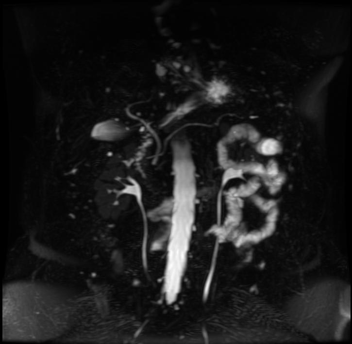
   Caption        = MRCP: Pancreas divisum. <br> [http://www.radswiki.net Image courtesy of RadsWiki]|
  DiseasesDB    = 31894 |
 
  ICD10          = {{ICD10|Q|45|3|q|38}}  |
  ICD9          = {{ICD9|751.7}} |
  ICDO          = |
  OMIM          = |
  MedlinePlus    = 000247 |
  eMedicineSubj  = radio |
  eMedicineTopic = 520 |
  MeshID        = |
}}
}}
{{SI}}
{{Pancreas divisum}}
{{CMG}}
'''For patient information on this topic, click [[{{PAGENAME}} (patient information)|here]]'''.


'''Contributors:'''  [[User:zorkun|Cafer Zorkun]] M.D., PhD.
{{CMG}}; {{AE}} [[User:zorkun|Cafer Zorkun]] M.D., PhD.


{{Editor Help}}
==[[Pancreas divisum overview|Overview]]==


==Overview==
==[[Pancreas divisum historical perspective|Historical Perspective]]==
'''Pancreas''' or '''Pancreatic divisum''' is a congenital anomaly in the [[anatomy]] of the ducts of the [[pancreas]] in which a single [[pancreatic duct]] is not formed, but rather remains as two distinct dorsal and ventral ducts.


*Variant of the pancreatic ductal system and occurs in 4%–10% of the population.
==[[Pancreas divisum pathophysiology|Pathophysiology]]==
*Dorsal and ventral anlagen of the pancreas fail to fuse.
**The dorsal duct drains most of the glandular parenchyma through the minor papilla
**The smaller ventral duct drains a portion of the pancreatic head, including the uncinate process, through the major papilla.
*Clinical relevance of pancreas divisum remains controversial.
**Most patients with pancreas divisum are asymptomatic.
**In some patients, this anomaly is associated with recurrent episodes of [[pancreatitis]].
**Postulated that in pancreas divisum, the duct of Santorini and the minor ampulla are too small to adequately drain the secretions produced by the pancreatic body and tail.


==Causes==
==[[Pancreas divisum causes|Causes]]==


The human embryo begins life with two ducts in the [[pancreas]], these are the ventral duct and the dorsal duct. Normally, the two ducts will fuse together to form one main pancreatic duct; this occurs in more than 90% of embryos. In approximately 10% of embryos the ventral and dorsal ducts fail to fuse together, resulting in pancreas divisum. In utero, the majority of the pancreas is drained by the dorsal duct which opens up into the minor papilla. The ventral duct drains the minority of the pancreas and opens into the major papilla. In adults however, this situation is reversed whereby 70% of the pancreas is drained by the ventral duct. Therefore in pancreas divisum, where fusion of the ducts does not occur, the major drainage of the pancreas is done by the dorsal duct which opens up into the minor papilla.
==[[Pancreas divisum differential diagnosis|Differentiating Pancreas Divisum from other Diseases]]==


[[Image:Pancreas_embryology.PNG]]
==[[Pancreas divisum epidemiology and demographics|Epidemiology and Demographics]]==


This is the embryologic progression of the pancreas formation. The 4<sup>th</sup> picture is a normal pancreas. The duct of santorini or minor papilla was closed and the dorsal pancreas drains into the ventral duct. In pancreas divisum, the last step (4<sup>th</sup> picture) does not happen. The pancreas formation is halted at the 3rd picture.
==[[Pancreas divisum risk factors|Risk Factors]]==


==Symptoms==
==[[Pancreas divisum natural history, complications and prognosis|Natural History, Complications and Prognosis]]==
 
A majority of individuals born with pancreas divisum will never have symptoms for their entire life. In most cases, pancreas divisum is only detected during an autopsy of a person that is deceased. However, approximately 1% of those with pancreas divisum will develop symptoms during their lifetime. Symptoms commonly include [[abdominal pain]], [[nausea]] and/or [[vomiting]], and [[pancreatitis]]. A small number of individuals may develop chronic pancreatitis.


==Diagnosis==
==Diagnosis==


The most common and accurate way of diagnosing an individual with this anomaly is by an [[ERCP]]. This test can demonstrate the presence of two separately draining ducts within the pancreas. Other tests can assist doctors with diagnosis, such as a [[CT scan]] and an [[MRI]].
[[Pancreas divisum history and symptoms|History and Symptoms]] | [[Pancreas divisum physical examination|Physical Examination]] | [[Pancreas divisum laboratory findings|Laboratory Findings]] | [[Pancreas divisum abdominal x ray|Abdominal X Ray]] | [[Pancreas divisum CT|CT]] | [[Pancreas divisum MRI|MRI]] | [[Pancreas divisum ultrasound|Ultrasound]] | [[Pancreas divisum other imaging findings|Other Imaging Findings]] | [[Pancreas divisum other diagnostic studies|Other Diagnostic Studies]]


==Diagnostic Findings==
==Treatment==
 
* The dorsal duct drains most of the glandular parenchyma through the minor papilla
* The smaller ventral duct drains a portion of the pancreatic head, including the uncinate process, through the major papilla.


'''Patient #1'''
[[Pancreas divisum medical therapy|Medical Therapy]] | [[Pancreas divisum surgery|Surgery]] | [[Pancreas divisum prevention|Prevention]] | [[Pancreas divisum cost-effectiveness of therapy|Cost-Effectiveness of Therapy]] | [[Pancreas divisum future or investigational therapies|Future or Investigational Therapies]]


[http://www.radswiki.net Images courtesy of RadsWiki]
==Case Studies==


<gallery>
[[Pancreas divisum case study one|Case #1]]
Image:Pancreatic-divisum-001.jpg
==Related Chapters==
Image:Pancreatic-divisum-002.jpg
Image:Pancreatic-divisum-003.jpg
</gallery>


'''Patient #2'''
*[[Anomalous pancreaticobiliary junction]]
*[[Annular pancreas]]


[http://www.radswiki.net Images courtesy of RadsWiki]
==External Links==


<gallery>
* [http://goldminer.arrs.org/search.php?query=Pancreatic%20divisum Goldminer: Pancreatic divisum]
Image:Pancreatic-divisum-101.jpg|MRCP
</gallery>
 
==Treatment==
 
Pancreas Divisum in individuals with no symptoms do not need treatment. Treatment of those with symptoms varies and has not been very well established. A surgeon may attempt a [[sphincterotomy]] by cutting the minor papilla to enlarge the opening and allow pancreas [[enzymes]] to flow normally. During surgery, a stent may be inserted into the duct to ensure that the duct will not close causing a blockage. As with any surgery, this operation is not risk-free. This surgery can cause pancreatitis in patients, or in rare cases, kidney failure and death.
 
An association with [[adenoma]] of the minor papilla has been reported.<ref name="pmid18019730">{{cite journal |author=Nakamura Y, Tajiri T, Uchida E, ''et al'' |title=Adenoma of the minor papilla associated with pancreas divisum |journal=Hepatogastroenterology |volume=54 |issue=78 |pages=1841–3 |year=2007 |pmid=18019730 |doi=}}</ref>
 
==References==
{{reflist}}
 
==External links==
* [http://www.medicinenet.com/pancreas_divisum/article.htm MedicineNet]
* {{Chorus|00303}}
* {{Chorus|00303}}


{{Congenital malformations and deformations of digestive system}}
{{SIB}}   


[[Category:Congenital disorders]]
[[Category:Congenital disorders]]
[[Category:Gastroenterology]]
[[Category:Disease]]
[[de:Pankreas divisum]]
[[de:Pankreas divisum]]


Latest revision as of 13:15, 11 April 2013

Pancreas divisum
MRCP: Pancreas divisum.
Image courtesy of RadsWiki

Pancreas Divisum Microchapters

Home

Patient Information

Overview

Historical Perspective

Pathophysiology

Causes

Differentiating Pancreas Divisum from other Diseases

Epidemiology and Demographics

Risk Factors

Natural History, Complications and Prognosis

Diagnosis

History and Symptoms

Physical Examination

Laboratory Findings

Abdominal X Ray

CT

MRI

Ultrasound

Other Imaging Findings

Other Diagnostic Studies

Treatment

Medical Therapy

Surgery

Prevention

Cost-Effectiveness of Therapy

Future or Investigational Therapies

Case Studies

Case #1

Pancreas divisum On the Web

Most recent articles

Most cited articles

Review articles

CME Programs

Powerpoint slides

Images

American Roentgen Ray Society Images of Pancreas divisum

All Images
X-rays
Echo & Ultrasound
CT Images
MRI

Ongoing Trials at Clinical Trials.gov

US National Guidelines Clearinghouse

NICE Guidance

FDA on Pancreas divisum

CDC on Pancreas divisum

Pancreas divisum in the news

Blogs on Pancreas divisum

Directions to Hospitals Treating Pancreas divisum

Risk calculators and risk factors for Pancreas divisum

For patient information on this topic, click here.

Editor-In-Chief: C. Michael Gibson, M.S., M.D. [1]; Associate Editor(s)-in-Chief: Cafer Zorkun M.D., PhD.

Overview

Historical Perspective

Pathophysiology

Causes

Differentiating Pancreas Divisum from other Diseases

Epidemiology and Demographics

Risk Factors

Natural History, Complications and Prognosis

Diagnosis

History and Symptoms | Physical Examination | Laboratory Findings | Abdominal X Ray | CT | MRI | Ultrasound | Other Imaging Findings | Other Diagnostic Studies

Treatment

Medical Therapy | Surgery | Prevention | Cost-Effectiveness of Therapy | Future or Investigational Therapies

Case Studies

Case #1

Related Chapters

External Links

Template:Congenital malformations and deformations of digestive system

de:Pankreas divisum


Template:WikiDoc Sources