Osteosarcoma x ray: Difference between revisions
Jump to navigation
Jump to search
No edit summary |
(→X Ray) |
||
| Line 4: | Line 4: | ||
==Overview== | ==Overview== | ||
==X Ray== | ==X Ray== | ||
Conventional radiography continues to play an important role in diagnosis. Typical appearances of conventional high grade osteosarcoma include: | Conventional radiography continues to play an important role in diagnosis. Typical appearances of conventional high grade osteosarcoma include:<ref name=radio2> Osteosarcoma. Dr Amir Rezaee ◉ and Dr Frank Gaillard ◉ et al. Radiopaedia.org 2015. http://radiopaedia.org/articles/osteosarcoma</ref> | ||
*Medullary and cortical bone destruction. | *Medullary and cortical bone destruction. | ||
*Wide zone of transition, permeative or moth-eaten appearance. | *Wide zone of transition, permeative or moth-eaten appearance. | ||
| Line 11: | Line 11: | ||
:*[[Codman triangle]] | :*[[Codman triangle]] | ||
:*Lamellated (onion skin) reaction: less frequently seen | :*Lamellated (onion skin) reaction: less frequently seen | ||
*Soft-tissue mass | *Soft-tissue mass. | ||
*Tumor matrix ossification/[[calcification]]. | *Tumor matrix ossification/[[calcification]]. | ||
:*Variable: reflects a combination of the amount of tumor bone production, calcified matrix, and [[osteoid]]. | :*Variable: reflects a combination of the amount of tumor bone production, calcified matrix, and [[osteoid]]. | ||
Revision as of 14:11, 28 September 2015
Editor-In-Chief: C. Michael Gibson, M.S., M.D. [1]
|
Osteosarcoma Microchapters |
|
Diagnosis |
|---|
|
Treatment |
|
Case Studies |
|
Osteosarcoma x ray On the Web |
|
American Roentgen Ray Society Images of Osteosarcoma x ray |
Overview
X Ray
Conventional radiography continues to play an important role in diagnosis. Typical appearances of conventional high grade osteosarcoma include:[1]
- Medullary and cortical bone destruction.
- Wide zone of transition, permeative or moth-eaten appearance.
- Aggressive periosteal reaction characterized by:
- Sunburst appearance
- Codman triangle
- Lamellated (onion skin) reaction: less frequently seen
- Soft-tissue mass.
- Tumor matrix ossification/calcification.
- Variable: reflects a combination of the amount of tumor bone production, calcified matrix, and osteoid.
- Ill-defined fluffy or cloud-like cf. to the rings and arcs of chondroid lesions.
-
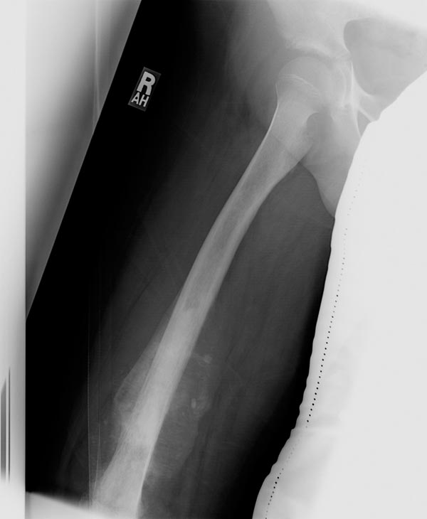
Plain film: Osteosarcoma
-
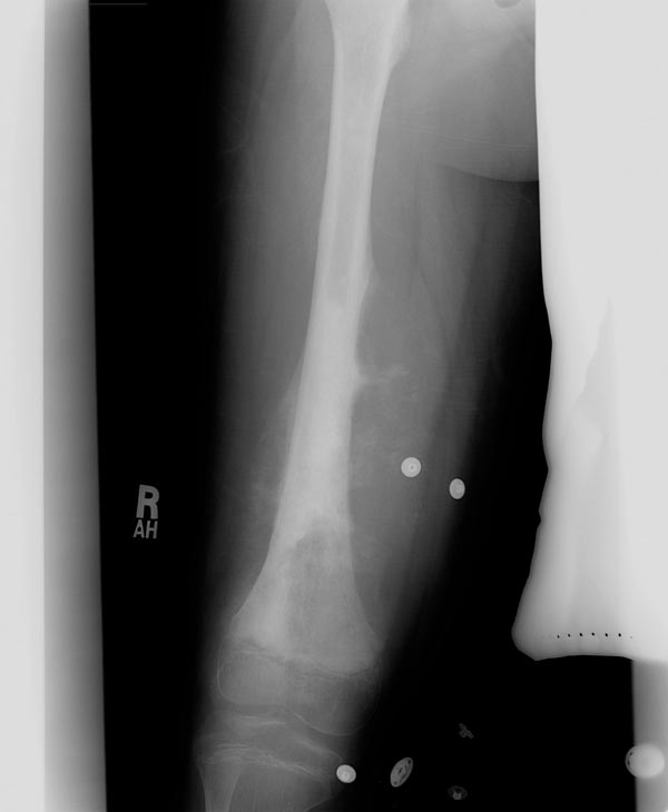
Plain film: Osteosarcoma


References
- ↑ Osteosarcoma. Dr Amir Rezaee ◉ and Dr Frank Gaillard ◉ et al. Radiopaedia.org 2015. http://radiopaedia.org/articles/osteosarcoma