Osteomyelitis MRI: Difference between revisions
Jump to navigation
Jump to search
(Created page with "__NOTOC__ {{Osteomyelitis}} Please help WikiDoc by adding content here. It's easy! Click here to learn about editing. ==References== {{Reflis...") |
No edit summary |
||
| Line 1: | Line 1: | ||
__NOTOC__ | __NOTOC__ | ||
{{Osteomyelitis}} | {{Osteomyelitis}} | ||
{{CMG}} | |||
Please help WikiDoc by adding content here. It's easy! Click [[Help:How_to_Edit_a_Page|here]] to learn about editing. | Please help WikiDoc by adding content here. It's easy! Click [[Help:How_to_Edit_a_Page|here]] to learn about editing. | ||
==Overview== | |||
Although MR imaging is the accepted modality of choice for the early detection and surgical localization of osteomyelitis, in the emergency department, CT is usually more readily available for establishing the diagnosis. <ref>Laura M. Fayad, John A. Carrino, and Elliot K. Fishman. [http://radiographics.rsnajnls.org/cgi/content/abstract/27/6/1723 Musculoskeletal Infection: Role of CT in the Emergency Department.] RadioGraphics 2007 27: 1723-1736.</ref> | |||
==MRI== | |||
[http://www.radswiki.net Images courtesy of RadsWiki] | |||
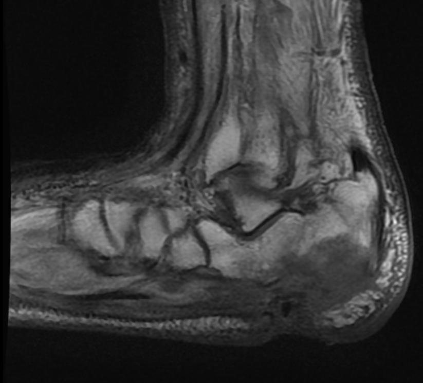
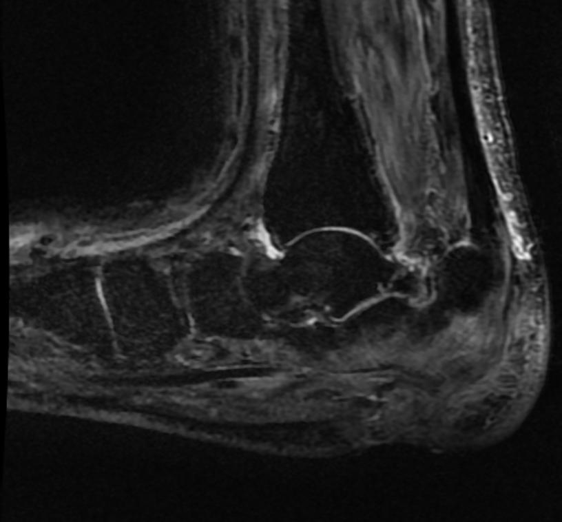
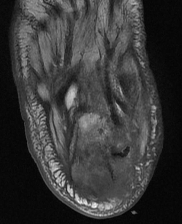
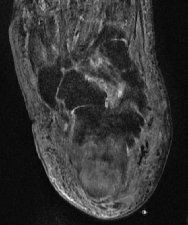
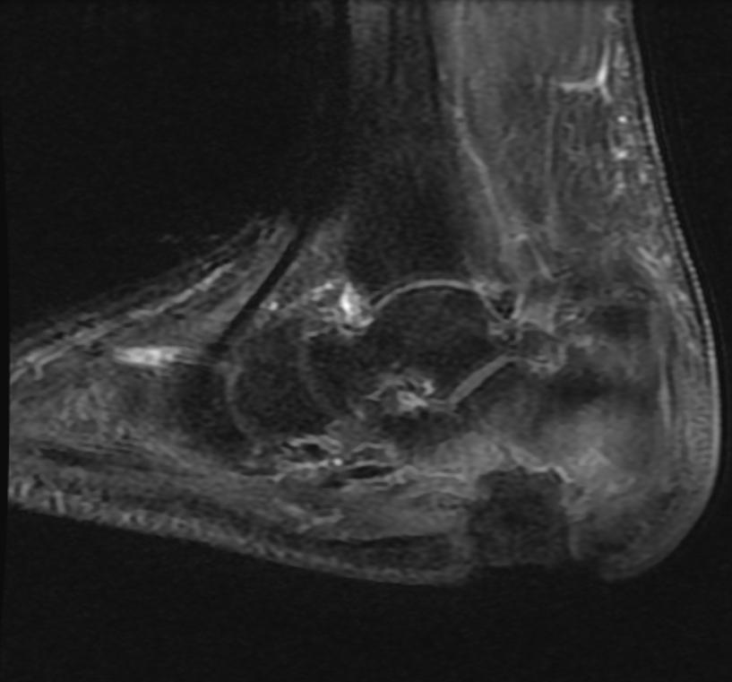
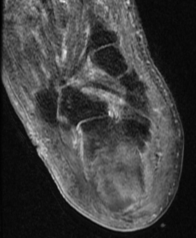
'''Patient #1 Extensive calcaneal osteomyelitis. Note soft tissue ulceration and [[cellulitis]]''' | |||
<gallery> | |||
Image:Osteomyelitis MRI 001.jpg|T1 | |||
Image:Osteomyelitis MRI 002.jpg|STIR | |||
Image:Osteomyelitis MRI 003.jpg|T1 | |||
Image:Osteomyelitis MRI 004.jpg|STIR | |||
Image:Osteomyelitis MRI 005.jpg|T1 fat sat contrast | |||
Image:Osteomyelitis MRI 006.jpg|T1 fat sat contrast | |||
</gallery> | |||
==References== | ==References== | ||
Revision as of 19:28, 19 December 2012
|
Osteomyelitis Microchapters |
|
Diagnosis |
|---|
|
Treatment |
|
Case Studies |
|
Osteomyelitis MRI On the Web |
|
American Roentgen Ray Society Images of Osteomyelitis MRI |
Editor-In-Chief: C. Michael Gibson, M.S., M.D. [1]
Please help WikiDoc by adding content here. It's easy! Click here to learn about editing.
Overview
Although MR imaging is the accepted modality of choice for the early detection and surgical localization of osteomyelitis, in the emergency department, CT is usually more readily available for establishing the diagnosis. [1]
MRI
Patient #1 Extensive calcaneal osteomyelitis. Note soft tissue ulceration and cellulitis
-
T1
-
STIR
-
T1
-
STIR
-
T1 fat sat contrast
-
T1 fat sat contrast


References
- ↑ Laura M. Fayad, John A. Carrino, and Elliot K. Fishman. Musculoskeletal Infection: Role of CT in the Emergency Department. RadioGraphics 2007 27: 1723-1736.