Orbit (anatomy)
Template:Infobox Anatomy Editor-In-Chief: C. Michael Gibson, M.S., M.D. [1]
Overview
In anatomy, the orbital bone is the cavity or socket of the skull in which the eye and its appendages are situated.
It can also mean the skin which surrounds the eye of a bird.
In the adult human, the volume of the orbit is 30 ml, of which the eye occupies 6.5 ml. [1]
Definition
The orbits are conical cavities, which open into the midline of the face. Each consists of a base, an apex and four walls.
The base, which opens in the face, has four borders. The following bones take part in their formation:
- 1. Superior margin: frontal bone
- 2. Inferior margin: maxilla and zygomatic
- 3. Medial margin: frontal, lacrimal and maxilla
- 4. Lateral margin: zygomatic and frontal
The apex lies near the medial end of superior orbital fissure and contain the optic canal which communicates with middle cranial fossa.
The roof (superior wall) is formed by the orbital plate frontal bone and the lesser wing of sphenoid. The orbital surface presents medially by trochlear fovea and laterally by lacrimal fossa
The floor (inferior wall) is formed by the orbital surface of maxilla, the orbital surface of zygomatic bone and the orbital process of palatine bone. Medially near the orbital margin is located the groove for nasolacrimal duct. Near the middle of the floor, located infraorbital groove, which leads to the infraorbital foramen. The floor is separated from the lateral wall by inferior orbital fissure, which connects the orbit to pterygopalatine and infratemporal fossa.
The medial wall is formed by the frontal process of maxilla, lacrimal bone, orbital plate of ethmoid and a small part of the body of the sphenoid.
The Lateral wall is formed by the orbital process of zygomatic and the orbital plate of greater wing of sphenoid. The bones meet at the zygomaticosphenoid suture. The lateral wall is the thickest wall of the orbit.
Fat cushion
In the orbit, surrounding the eyeball and its muscles, is a layer of fat that helps the eye rotate around a fixed center of rotation. If excess liquid is collected in the fat cushion tissue, the eye may protrude. [2]
Contents
- Eyeball
- Fascias: Orbital, Bulbar
- Extraocular muscles (Levator Palpebrae Superioris, Superior, Inferior, Lateral and Medial Rectus muscles, Superior and Inferior Oblique Muscles)
- Nerves: cranial nerves II, III, IV, V, and VI
- Blood vessels
- Extraocular Fat
- Lacrimal gland, Lacrimal sac, Nasolacrimal duct
- Eyelids
- Medial and Lateral Palpebral ligaments
- Medial and Lateral Check ligaments
- Suspensory ligament of the eyeball
- Conjunctiva
- Trochlea
- Orbital septum
- Ciliary ganglion and short ciliary nerves
Bones
In humans, seven bones make up the bony orbit:
- Frontal bone (Pars orbitalis)
- Lacrimal bone
- Ethmoid bone (Lamina papyracea)
- Zygomatic bone (Orbital process of the the zygomatic bone)
- Maxillary bone (Orbital surface of the body of the maxilla)
- Palatine bone (Orbital process of palatine bone)
- Sphenoid bone (Greater and lesser wings)
Foramina and openings
- Optic foramen
- Superior orbital fissure
- Inferior orbital fissure
- Anterior ethmoid foramen
- Posterior ethmoidal foramen
- Infraorbital foramen
- Supraorbital foramen
- Naso-lacrimal canal opening
- Zygomatic orbital foramen
Additional images
-

The skull from the front.
-

Medial wall of left orbit.
-
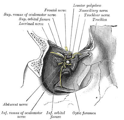
Dissection showing origins of right ocular muscles, and nerves entering by the superior orbital fissure.
-

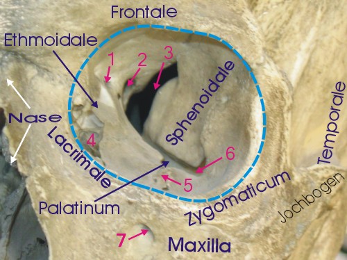
Coronal section of nasal cavities.
-
-
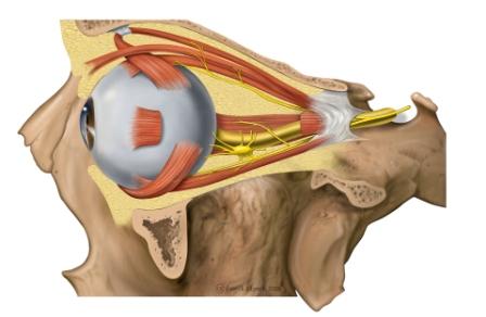
Lateral orbit anatomy
-
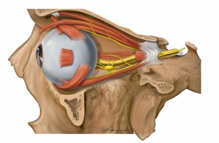
Lateral orbit nerves


References
External links
- oph/2 at eMedicine - "Arterial Supply, Orbit"
- Template:SUNYAnatomyLabs
- Template:UMichAtlas
- Template:UMichAtlas
- Interactive tutorial at anatome.ncl.ac.uk
de:Orbita
it:Orbita oculare
la:Orbita (anatomia)
nl:Orbita
fi:Silmäkuoppa
sv:Ögonhåla