Onychomycosis physical examination: Difference between revisions
Shanshan Cen (talk | contribs) No edit summary |
m (Changes made per Mahshid's request) |
||
| Line 41: | Line 41: | ||
[[Category:Disease]] | [[Category:Disease]] | ||
Revision as of 18:32, 18 September 2017
|
Onychomycosis Microchapters |
|
Diagnosis |
|---|
|
Treatment |
|
Case Studies |
|
Onychomycosis physical examination On the Web |
|
American Roentgen Ray Society Images of Onychomycosis physical examination |
|
Risk calculators and risk factors for Onychomycosis physical examination |
Editor-In-Chief: C. Michael Gibson, M.S., M.D. [1]
Physical Examination
The following images show examples of how an onychomycosis patient's nails may look like. The nails may have a brown appearance.
(Images courtesy of Charlie Goldberg, M.D., UCSD School of Medicine and VA Medical Center, San Diego, CA)
-
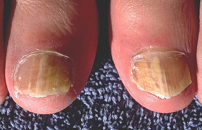
Onychomycosis due to Trychophyton rubrum, right and left great toe.
-
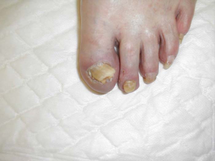
Onychomycosis: Chronic fungal infection causing discoloration and deformity of nails.
-
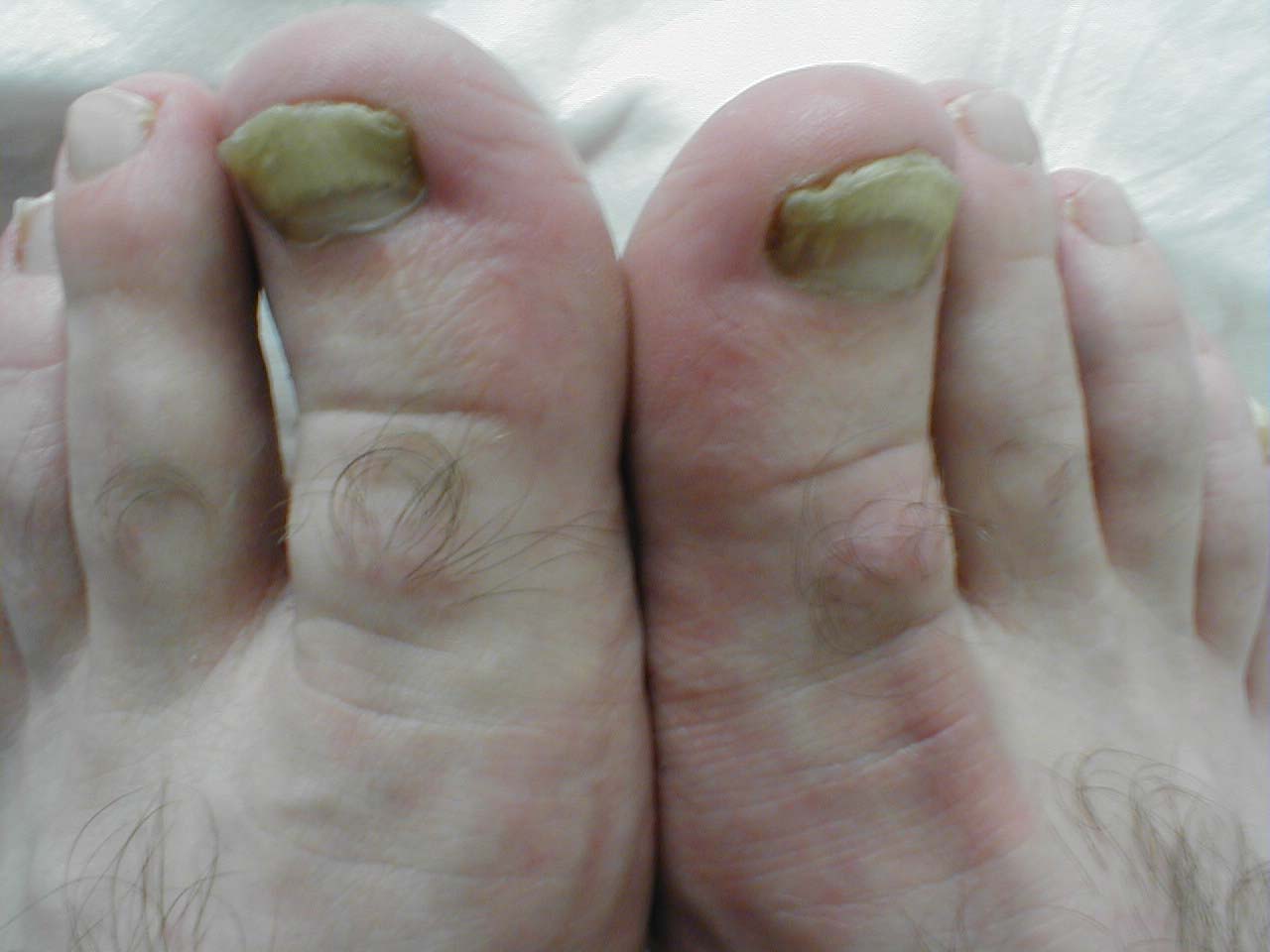
Onychomycosis: Chronic fungal toenail infection causing deformity and discoloration.


Nails
-
![Onychomycosis. With permission from Dermatology Atlas.[1]](/images/c/ca/Onychomycosis01.jpg)
Onychomycosis. With permission from Dermatology Atlas.[1]
-
![Onychomycosis. With permission from Dermatology Atlas.[1]](/images/5/58/Onychomycosis02.jpg)
Onychomycosis. With permission from Dermatology Atlas.[1]
-
![Onychomycosis. With permission from Dermatology Atlas.[1]](/images/0/05/Onychomycosis03.jpg)
Onychomycosis. With permission from Dermatology Atlas.[1]
-
![Onychomycosis. With permission from Dermatology Atlas.[1]](/images/1/1e/Onychomycosis04.jpg)
Onychomycosis. With permission from Dermatology Atlas.[1]
![Onychomycosis. With permission from Dermatology Atlas.[1]](/index.php/File:Onychomycosis01.jpg)
![Onychomycosis. With permission from Dermatology Atlas.[1]](/index.php/File:Onychomycosis02.jpg)
![Onychomycosis. With permission from Dermatology Atlas.[1]](/index.php/File:Onychomycosis03.jpg)
![Onychomycosis. With permission from Dermatology Atlas.[1]](/index.php/File:Onychomycosis04.jpg)