Oligodendroglioma MRI: Difference between revisions
Jump to navigation
Jump to search
| Line 9: | Line 9: | ||
===Gallery=== | ===Gallery=== | ||
<gallery> | <gallery> | ||
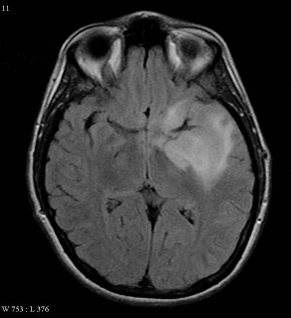
Image:Oligodendroglioma axial FLAIR.jpg|A relatively well circumscribed mass involves the temporal lobe and insular cortex, without convincing enhancement, and minimal restricted diffusion.<ref name=MRI radio>Image courtesy of Dr. Frank Gaillard. Radiopaedia (original file [http://radiopaedia.org/cases/oligodendroglioma-9 here]). Creative Commons BY-SA-NC</ref> | |||
</gallery> | </gallery> | ||
Revision as of 15:12, 8 October 2015
|
Oligodendroglioma Microchapters |
|
Diagnosis |
|---|
|
Treatment |
|
Case Studies |
|
Oligodendroglioma MRI On the Web |
|
American Roentgen Ray Society Images of Oligodendroglioma MRI |
Editor-In-Chief: C. Michael Gibson, M.S., M.D. [1]Associate Editor(s)-in-Chief: Sujit Routray, M.D. [2]
Overview
MRI
Gallery
-
A relatively well circumscribed mass involves the temporal lobe and insular cortex, without convincing enhancement, and minimal restricted diffusion.
