Notochord
|
WikiDoc Resources for Notochord |
|
Articles |
|---|
|
Most recent articles on Notochord |
|
Media |
|
Evidence Based Medicine |
|
Clinical Trials |
|
Ongoing Trials on Notochord at Clinical Trials.gov Clinical Trials on Notochord at Google
|
|
Guidelines / Policies / Govt |
|
US National Guidelines Clearinghouse on Notochord
|
|
Books |
|
News |
|
Commentary |
|
Definitions |
|
Patient Resources / Community |
|
Patient resources on Notochord Discussion groups on Notochord Directions to Hospitals Treating Notochord Risk calculators and risk factors for Notochord
|
|
Healthcare Provider Resources |
|
Causes & Risk Factors for Notochord |
|
Continuing Medical Education (CME) |
|
International |
|
|
|
Business |
|
Experimental / Informatics |
Overview
The notochord is a flexible, rod-shaped body found in embryos of all chordates. It is composed of cells derived from the mesoderm and defines the primitive axis of the embryo. In lower vertebrates, it persists throughout life as the main axial support of the body, while in higher vertebrates it is replaced by the vertebral column. The notochord is found on the ventral surface of the neural tube.
Notochords were the first "backbones", as well, serving as support structures in chordates that lacked a bony skeleton. The very first vertebrates, such as Haikouicthys, had only a notochord. Embryos of vertebrates have notochords today, as embryonic development often happens to follow a pattern similar to the ancestral evolution of the modern animal's traits [this idea that 'ontogeny recapitulates phylogeny' is something of an old-fashioned concept in embryology]. Notochords were advantageous to primitive fish-ancestors because they were a rigid structure for muscle attachment, yet flexible enough to allow more movement than, for example, the exoskeleton of the dominant animals of that time. In humans, they eventually develop into the disks between the vertebrae.
Development
Notogenesis is the development of the notochord by the epiblasts that make up the floor of the amnion cavity (Human Embryology). The notochord arises as a pouch from the mesoderm.
The notochord forms during gastrulation and soon after induces the formation of the neural plate (neurulation), synchronizing the development of the neural tube. On the ventral aspect of the neural groove an axial thickening of the endoderm takes place. (In bi-pedal chordates, e.g. humans, this surface is properly referred to as the anterior surface). This thickening appears as a furrow (the chordal furrow) the margins of which anastimose (come into contact), and so convert it into a solid rod of polygonal-shaped cells (the notochord) which is then separated from the endoderm.
In higher vertebrates, it extends throughout the entire length of the future vertebral column, and reaches as far as the anterior end of the midbrain, where it ends in a hook-like extremity in the region of the future dorsum sellæ of the sphenoid bone. Initially it exists between the neural tube and the endoderm of the yolk-sac, but soon becomes separated from them by the mesoderm, which grows medially and surrounds it. From the mesoderm surrounding the neural tube and notochord, the skull, vertebral column, and the membranes of the brain and medulla spinalis are developed.
Postembryonic vestige of the notochord is found in the nucleus pulposus of the intervertebral disks, but not in the vertebral bodies, from which notochordal cells usually regress entirely. In humans, by the age of 4, all notochord residue is replaced by a population of chondrocyte-like cells of unclear origin[1]. Persistence of notochordal cells within the vertebra may cause a pathologic condition- persistent notochordal canal[2]. They are also found to persist in the nasopharyngeal space and, in such an unusual instance, may give rise to Tornwaldt's cyst.
Research
Research into the notochord has played a key role in understanding the development of the central nervous system. By transplanting and expressing a second notochord near the dorsal neural tube, 180 degrees opposite of the normal notochord location, one can induce the formation of motoneurons in the dorsal tube. Motoneuron formation generally occurs in the ventral neural tube, while the dorsal tube generally forms sensory cells.
The notochord secretes a protein called sonic hedgehog homolog (SHH), a key morphogen regulating organogenesis and having a critical role in signaling the development of motoneurons[3]. The secretion of SHH by the notochord establishes the ventral pole of the dorsal-ventral axis in the developing embryo.
Additional images
-
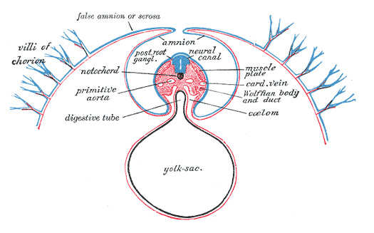
Diagram of a transverse section, showing the mode of formation of the amnion in the chick.
-
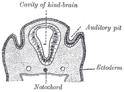
Section through the head of a human embryo, about twelve days old, in the region of the hind-brain.
-
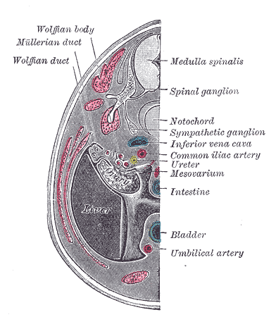
Transverse section of human embryo eight and a half to nine weeks old.


Reference
- ↑ J. P. G. Urban , S. Roberts , and J. R. Ralphs. The Nucleus of the Intervertebral Disc from Development to Degeneration. Amer Zool. 2000;40(1):53-61. full text
- ↑ Christopherson LR, Rabin BM, Hallam DK, Russell EJ. Persistence of the notochordal canal: MR and plain film appearance. AJNR Am J Neuroradiol. 1999 Jan;20(1):33-6. PMID 9974055
- ↑ Echelard Y, Epstein DJ, St-Jacques B, Shen L, Mohler J, McMahon JA, McMahon AP. Sonic hedgehog, a member of a family of putative signaling molecules, is implicated in the regulation of CNS polarity. Cell 1993;75(7):1417-30. PMID 7916661
External links
Template:Development of nervous system
ca:Notocordi cs:Struna hřbetní de:Chorda dorsalis eo:Notokordo it:Notocorda