Non ST Elevation Myocardial Infarction: Diagnosis: Difference between revisions

Jump to navigation Jump to search
No edit summary
 
m (Robot: Automated text replacement (-{{WikiDoc Cardiology Network Infobox}} +, -<references /> +{{reflist|2}}, -{{reflist}} +{{reflist|2}}))
 
(2 intermediate revisions by 2 users not shown)
Line 1: Line 1:
[[image:unstable-angina.jpg|thumb|220px|ST Depression in a pt. with unstable angina]]
[[image:unstable-angina.jpg|thumb|220px|ST Depression in a pt. with unstable angina]]
{{SI}}
{{SI}}
{{WikiDoc Cardiology Network Infobox}}
 
{{WikiDoc Cardiology News}}
{{CMG}}
{{CMG}}
Line 7: Line 7:
'''Associate Editor-In-Chief:''' {{CZ}}


{{Editor Join}}


== History and Symptoms ==


A person with unstable angina (UA) will have a history of angina that has increased in frequency or intensity at the same level of exertion. Anginal pain can manifest in many forms ranging from chest pain to chest pressure to shortness of breath to epigastric pain. As mentioned above, UA is in the spectrum of ACS and requires immediate assessment by a qualified physician. The history and symptoms described by a patient with NSTEMI can be identical to those of the patient presenting with a STEMI and thus it is most useful to describe the classic history and symptoms suggestive of ACS.


According to the ACC/AHA UA/NSTEMI guidelines, most important factors on the initial history are
1) the nature of the anginal symptoms,
2) prior history of CAD (e.g., prior myocardial infarction (MI), angina, cardiac catheterization, coronary artery bipass grafting (CABG)),
3) male gender,
4) older age
5) an increasing number of traditional risk factors. <cite>andersonref1</cite>  Other pertinent medical history which will help with risk-stratification should also be obtained rapidly, including cardiac risk factors (i.e., family history of premature coronary artery disease in a first degree relative < 60 yrs old, elevated cholesterol, hypertension, diabetes mellitus, smoking history past or present), current medications and allergies.<cite>giblerref1</cite>
The most common history given by a patient with ACS is that of chest discomfort, described as crushing, left-sided substernal chest pain or pressure that radiates to the neck or left arm. Indeed, sometimes it is described as the sensation of "an elephant is sitting on the chest."  However, in reality the history can be quite variable. The pain is sometimes located solely in the epigastric region, the right side of the chest, the jaw, neck, arm, shoulder or back and a history of nausea, dyspnea or diaphoresis is not infrequent. <cite>andersonref1</cite> It is important to note that certain patient populations may be even less likely to present with classic symptoms. These groups include women, older patients and patients with renal failure and diabetes. <cite>giblerref1</cite> Pleuritic pain (sharp pain on inspiration or from a cough), mid/lower abdominal pain, pain reproducible with palpation or movement, very brief episodes of pain (e.g., seconds) and pain that radiates to the lower extremities are all traits that are less likely to be from - although they do not exclude - ACS. Similarly, a history that nitroglycerine does not relieve the pain or a history that a "GI coctail" does relieve the pain is less suggestive of ACS, although ACS still cannot be excluded on this basis. <cite>andersonref1</cite>
A thorough history of present illness (HPI) obtained by the physician will include the time of onset, duration, location, radiation, quality, intensity, aggravating and relieving factors (i.e., deep breathing, position, exertion), associated symptoms (i.e., diaphoresis, nausea, vomiting, dyspnea, dizziness), any history of prior similar symptoms along with a comparison of the pain to any previously diagnosed angina.<cite>giblerref1</cite>


== Laboratory Findings ==  
Line 80: Line 62:
* [[The Living Guidelines: UA/NSTEMI]]


{{SIB}}
 
[[Category:Cardiology]]
[[Category:Cardiology]]
{{WikiDoc Help Menu}}
{{WikiDoc Sources}}
{{WikiDoc Sources}}
{{mdr}}
{{mdr}}

Latest revision as of 20:09, 4 September 2012
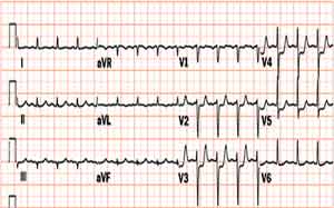
ST Depression in a pt. with unstable angina

WikiDoc Resources for Non ST Elevation Myocardial Infarction: Diagnosis

Articles

Most recent articles on Non ST Elevation Myocardial Infarction: Diagnosis

Most cited articles on Non ST Elevation Myocardial Infarction: Diagnosis

Review articles on Non ST Elevation Myocardial Infarction: Diagnosis

Articles on Non ST Elevation Myocardial Infarction: Diagnosis in N Eng J Med, Lancet, BMJ

Media

Powerpoint slides on Non ST Elevation Myocardial Infarction: Diagnosis

Images of Non ST Elevation Myocardial Infarction: Diagnosis

Photos of Non ST Elevation Myocardial Infarction: Diagnosis

Podcasts & MP3s on Non ST Elevation Myocardial Infarction: Diagnosis

Videos on Non ST Elevation Myocardial Infarction: Diagnosis

Evidence Based Medicine

Cochrane Collaboration on Non ST Elevation Myocardial Infarction: Diagnosis

Bandolier on Non ST Elevation Myocardial Infarction: Diagnosis

TRIP on Non ST Elevation Myocardial Infarction: Diagnosis

Clinical Trials

Ongoing Trials on Non ST Elevation Myocardial Infarction: Diagnosis at Clinical Trials.gov

Trial results on Non ST Elevation Myocardial Infarction: Diagnosis

Clinical Trials on Non ST Elevation Myocardial Infarction: Diagnosis at Google

Guidelines / Policies / Govt

US National Guidelines Clearinghouse on Non ST Elevation Myocardial Infarction: Diagnosis

NICE Guidance on Non ST Elevation Myocardial Infarction: Diagnosis

NHS PRODIGY Guidance

FDA on Non ST Elevation Myocardial Infarction: Diagnosis

CDC on Non ST Elevation Myocardial Infarction: Diagnosis

Books

Books on Non ST Elevation Myocardial Infarction: Diagnosis

News

Non ST Elevation Myocardial Infarction: Diagnosis in the news

Be alerted to news on Non ST Elevation Myocardial Infarction: Diagnosis

News trends on Non ST Elevation Myocardial Infarction: Diagnosis

Commentary

Blogs on Non ST Elevation Myocardial Infarction: Diagnosis

Definitions

Definitions of Non ST Elevation Myocardial Infarction: Diagnosis

Patient Resources / Community

Patient resources on Non ST Elevation Myocardial Infarction: Diagnosis

Discussion groups on Non ST Elevation Myocardial Infarction: Diagnosis

Patient Handouts on Non ST Elevation Myocardial Infarction: Diagnosis

Directions to Hospitals Treating Non ST Elevation Myocardial Infarction: Diagnosis

Risk calculators and risk factors for Non ST Elevation Myocardial Infarction: Diagnosis

Healthcare Provider Resources

Symptoms of Non ST Elevation Myocardial Infarction: Diagnosis

Causes & Risk Factors for Non ST Elevation Myocardial Infarction: Diagnosis

Diagnostic studies for Non ST Elevation Myocardial Infarction: Diagnosis

Treatment of Non ST Elevation Myocardial Infarction: Diagnosis

Continuing Medical Education (CME)

CME Programs on Non ST Elevation Myocardial Infarction: Diagnosis

International

Non ST Elevation Myocardial Infarction: Diagnosis en Espanol

Non ST Elevation Myocardial Infarction: Diagnosis en Francais

Business

Non ST Elevation Myocardial Infarction: Diagnosis in the Marketplace

Patents on Non ST Elevation Myocardial Infarction: Diagnosis

Experimental / Informatics

List of terms related to Non ST Elevation Myocardial Infarction: Diagnosis

Template:WikiDoc Cardiology News Editor-In-Chief: C. Michael Gibson, M.S., M.D. [1]

Associate Editor-In-Chief: Cafer Zorkun, M.D., Ph.D. [2]


Laboratory Findings

Electrolyte and Biomarker Studies

If there is an elevation of a marker of myocardial necrosis (CK-MB or troponin), then the patient does not have unstable angina, but instead has a syndrome of either ST elevation MI or Non ST elevation MI depending upon the EKG changes.

Electrocardiogram

ST Depression in a pt. with unstable angina

The resting electrocardiogram may show either

  1. No changes
  2. Flipped T waves
  3. ST Depression as shown to the right. ST depression carries the poorest prognosis.

Chest X Ray

A Chest X Ray is critical to aid in the exclusion of aortic dissection.

A mediastinal mass consistent with a cancer may be present, but it is unlikely to present with a syndrome of accelerating chest pain.

Differential Diagnosis of Chest Pain

Cardiovascular

References

  1. PMID 16046952
  2. PMID 17692756
  3. Bickley, LS (2003). Bates' Guide to Physical Examination and History Taking. Lippincott: Philadelphia, PA. ISBN 0781735114

See Also

Template:WikiDoc Sources Template:Mdr