Mydriasis: Difference between revisions
No edit summary |
No edit summary |
||
| Line 11: | Line 11: | ||
The mechanism of mydriasis depends on the agent being used. It usually involves either a disruption of the [[parasympathetic nerve]] supply to the eye (which causes contraction of the pupil), or over activity of the [[sympathetic nervous system]] (SNS). | The mechanism of mydriasis depends on the agent being used. It usually involves either a disruption of the [[parasympathetic nerve]] supply to the eye (which causes contraction of the pupil), or over activity of the [[sympathetic nervous system]] (SNS). | ||
==Causes== | ==Causes== | ||
===Common Causes=== | ===Common Causes=== | ||
===Non Pathological=== | ===Non Pathological=== | ||
Mydriasis can be congenital. | Mydriasis can be congenital. | ||
===Pathological=== | ===Pathological=== | ||
The [[parasympathetic]] nervous supply which causes constriction of the pupil, or [[miosis]], is supplied by [[cranial nerve]] III, the [[oculomotor nerve]]. Damage to this nerve typically manifests itself as mydriasis, because the [[sympathetic]] supply to the pupil which causes mydriasis remains unaffected, and therefore unopposed. | The [[parasympathetic]] nervous supply which causes constriction of the pupil, or [[miosis]], is supplied by [[cranial nerve]] III, the [[oculomotor nerve]]. Damage to this nerve typically manifests itself as mydriasis, because the [[sympathetic]] supply to the pupil which causes mydriasis remains unaffected, and therefore unopposed. | ||
===Traumatic=== | ===Traumatic=== | ||
In cases of [[head injury]] or [[eye injury|orbit trauma (eye injury)]], the [[Iris sphincter muscle|iris sphincter]] (the muscle responsible for closing the pupil) or the nerves controlling it can be damaged, reducing or eliminating [[reactivity]] to light. | In cases of [[head injury]] or [[eye injury|orbit trauma (eye injury)]], the [[Iris sphincter muscle|iris sphincter]] (the muscle responsible for closing the pupil) or the nerves controlling it can be damaged, reducing or eliminating [[reactivity]] to light. | ||
===Drugs=== | ===Drugs=== | ||
[[Atropine]] blocks [[muscarinic acetylcholine receptor]]s. [[Acetylcholine|Acetylcholine (ACh)]] is the [[neurotransmitter]] of the [[parasympathetic nervous system]] and blocking its action means the pupil cannot constrict. | [[Atropine]] blocks [[muscarinic acetylcholine receptor]]s. [[Acetylcholine|Acetylcholine (ACh)]] is the [[neurotransmitter]] of the [[parasympathetic nervous system]] and blocking its action means the pupil cannot constrict. | ||
| Line 32: | Line 27: | ||
[[Antihistamine]]s and [[tricyclic antidepressant]]s may cause mydriasis. | [[Antihistamine]]s and [[tricyclic antidepressant]]s may cause mydriasis. | ||
===Mydriatic Drops=== | ===Mydriatic Drops=== | ||
A '''mydriatic''' is an agent which induces [[dilation]] of the [[pupil]]. Drugs such as [[tropicamide]] are used in [[medicine]] to permit examination of the [[retina]] and other deep structures of the eye, and also to reduce painful [[ciliary muscle]] [[muscle spasm|spasm]] (see [[cycloplegia]]). One effect of administration of a mydriatic is intolerance to bright light. | A '''mydriatic''' is an agent which induces [[dilation]] of the [[pupil]]. Drugs such as [[tropicamide]] are used in [[medicine]] to permit examination of the [[retina]] and other deep structures of the eye, and also to reduce painful [[ciliary muscle]] [[muscle spasm|spasm]] (see [[cycloplegia]]). One effect of administration of a mydriatic is intolerance to bright light. | ||
===Physiological Response Indicating Interest=== | ===Physiological Response Indicating Interest=== | ||
[[Pupillary response]] may also indicate interest in the subject of attention or sexual stimulation.<ref>{{Citation | [[Pupillary response]] may also indicate interest in the subject of attention or sexual stimulation.<ref>{{Citation | ||
| last1 = Hess | first1 = Eckhard H. | | last1 = Hess | first1 = Eckhard H. | ||
| Line 66: | Line 57: | ||
*[[Trauma]] | *[[Trauma]] | ||
==Diagnostic Findings== | ==Diagnostic Findings== | ||
=== History and Symptoms === | === History and Symptoms === | ||
*Complete history with special attention to: | *Complete history with special attention to: | ||
:* | :*Neurologic | ||
:*Ophthalmologic | :*Ophthalmologic | ||
:*Otolaryngologic | :*Otolaryngologic | ||
=== Physical Examination === | === Physical Examination === | ||
=== Eyes === | === Eyes === | ||
<gallery> | |||
Image:Dilated_pupil.gk.jpg|Pupil dilated using [[anaesthetic]] and muscle relaxant | |||
Image:Eye dilate.gif|Pupillary response | |||
</gallery> | |||
*Pupil size (light & dark) | *Pupil size (light & dark) | ||
*Pupil response to light and convergence | *Pupil response to light and convergence | ||
*Lid position | *Lid position | ||
===MRI=== | ===MRI=== | ||
*MRI to check for [[third cranial nerve palsy]] | *MRI to check for [[third cranial nerve palsy]] | ||
==Treatment== | ==Treatment== | ||
===Medical Therapy=== | ===Medical Therapy=== | ||
*[[Adie's pupil]] - Pilcarpine .125% BID-QID | *[[Adie's pupil]] - Pilcarpine .125% BID-QID | ||
*Migraines - pain meds, antidepressants, anticonvulsants, beta blockers, calcium channel blockers | *Migraines - pain meds, antidepressants, anticonvulsants, beta blockers, calcium channel blockers | ||
===Primary Prevention=== | ===Primary Prevention=== | ||
*Remove causative medication | *Remove causative medication | ||
| Line 96: | Line 83: | ||
*[[Miosis]] | *[[Miosis]] | ||
*[[Anisocoria]] | *[[Anisocoria]] | ||
==References== | ==References== | ||
{{reflist|2}} | {{reflist|2}} | ||
Revision as of 21:25, 21 February 2013
|
WikiDoc Resources for Mydriasis |
|
Articles |
|---|
|
Most recent articles on Mydriasis |
|
Media |
|
Evidence Based Medicine |
|
Clinical Trials |
|
Ongoing Trials on Mydriasis at Clinical Trials.gov Clinical Trials on Mydriasis at Google
|
|
Guidelines / Policies / Govt |
|
US National Guidelines Clearinghouse on Mydriasis
|
|
Books |
|
News |
|
Commentary |
|
Definitions |
|
Patient Resources / Community |
|
Patient resources on Mydriasis Discussion groups on Mydriasis Directions to Hospitals Treating Mydriasis Risk calculators and risk factors for Mydriasis
|
|
Healthcare Provider Resources |
|
Causes & Risk Factors for Mydriasis |
|
Continuing Medical Education (CME) |
|
International |
|
|
|
Business |
|
Experimental / Informatics |
Editor-In-Chief: C. Michael Gibson, M.S., M.D. [2]
Overview
Mydriasis is an excessive dilation of the pupil due to disease, trauma or drugs. Normally, the pupil dilates in the dark and constricts in the light. A mydriatic pupil will remain excessively large, even in a bright environment. Sometimes colloquially referred to as a "blown pupil."
The opposite, constriction of the pupil, is called miosis.
Pathophysiology
There are two types of muscle that control the size of the iris: circular muscle and radial muscle. The former is innervated by the parasympathetic nervous system, the latter by the sympathetic nervous system. Sympathetic stimulation of α1 adrenergic receptors causes the contraction of the radial muscle, and subsequent dilation of the pupil. Conversely, parasympathetic stimulation cause contraction of the circular muscle and constriction of the iris.
The mechanism of mydriasis depends on the agent being used. It usually involves either a disruption of the parasympathetic nerve supply to the eye (which causes contraction of the pupil), or over activity of the sympathetic nervous system (SNS).
Causes
Common Causes
Non Pathological
Mydriasis can be congenital.
Pathological
The parasympathetic nervous supply which causes constriction of the pupil, or miosis, is supplied by cranial nerve III, the oculomotor nerve. Damage to this nerve typically manifests itself as mydriasis, because the sympathetic supply to the pupil which causes mydriasis remains unaffected, and therefore unopposed.
Traumatic
In cases of head injury or orbit trauma (eye injury), the iris sphincter (the muscle responsible for closing the pupil) or the nerves controlling it can be damaged, reducing or eliminating reactivity to light.
Drugs
Atropine blocks muscarinic acetylcholine receptors. Acetylcholine (ACh) is the neurotransmitter of the parasympathetic nervous system and blocking its action means the pupil cannot constrict.
Cocaine inhibits the reuptake of noradrenaline (norepinephrine) within a nerve synapse. When a solution of cocaine is dropped into the eye, noradrenaline is no longer reabsorbed by neurons, and its levels increase. Noradrenaline, the neurotransmitter for the SNS, causes dilation of the pupil. Mydriasis is used as a diagnostic test for Horner's Syndrome, in which it is initiated using a similar mechanism, though cocaine is not usually used in this procedure. Many other drugs such as amphetamines and psychedelic drugs (LSD, psychedelic mushrooms, mescaline, and MDMA) are also known to cause mydriasis.
Opiate withdrawal can cause dilated pupils in some people. [1]
Antihistamines and tricyclic antidepressants may cause mydriasis.
Mydriatic Drops
A mydriatic is an agent which induces dilation of the pupil. Drugs such as tropicamide are used in medicine to permit examination of the retina and other deep structures of the eye, and also to reduce painful ciliary muscle spasm (see cycloplegia). One effect of administration of a mydriatic is intolerance to bright light.
Physiological Response Indicating Interest
Pupillary response may also indicate interest in the subject of attention or sexual stimulation.[2]
Causes in Alphabetical Order[3][4]
- Acute Closed Angle glaucoma
- Adie's tonic pupil
- Adregenic agents
- Benign episodic mydriasis
- Coma
- Congenital mydriasis
- Drug withdrawal
- Hallucinogens
- Seizures
- Serotonin syndrome
- Systemic anticholinergics
- Third nerve palsy
- Trauma
Diagnostic Findings
History and Symptoms
- Complete history with special attention to:
- Neurologic
- Ophthalmologic
- Otolaryngologic
Physical Examination
Eyes
-

Pupil dilated using anaesthetic and muscle relaxant
-
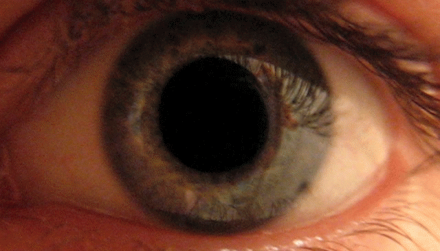
Pupillary response


- Pupil size (light & dark)
- Pupil response to light and convergence
- Lid position
MRI
- MRI to check for third cranial nerve palsy
Treatment
Medical Therapy
- Adie's pupil - Pilcarpine .125% BID-QID
- Migraines - pain meds, antidepressants, anticonvulsants, beta blockers, calcium channel blockers
Primary Prevention
- Remove causative medication
- Sunglasses to decrease light sensitivity
Related Chapters
References
- ↑ [1]
- ↑ Hess, Eckhard H.; Polt, James M. (5 August 1960), "Pupil Size as Related to Interest Value of Visual Stimuli", Science, 132 (3423): 349, doi:10.1126/science.132.3423.349
- ↑ Sailer, Christian, Wasner, Susanne. Differential Diagnosis Pocket. Hermosa Beach, CA: Borm Bruckmeir Publishing LLC, 2002:77 ISBN 1591032016
- ↑ Kahan, Scott, Smith, Ellen G. In A Page: Signs and Symptoms. Malden, Massachusetts: Blackwell Publishing, 2004:68 ISBN 140510368X