Multicystic dysplastic kidney
| Multicystic dysplastic kidney | |
 | |
|---|---|
| Ultrasonography: Multicystic dysplastic kidney Image courtesy of RadsWiki | |
| ICD-10 | Q61.4 (EUROCAT Q61.40-Q61.41) |
For patient information, click here
|
Multicystic dysplastic kidney Microchapters |
|
Differentiating Multicystic dysplastic kidney from other Diseases |
|---|
|
Diagnosis |
|
Treatment |
|
Case Studies |
|
Multicystic dysplastic kidney On the Web |
|
American Roentgen Ray Society Images of Multicystic dysplastic kidney |
|
Directions to Hospitals Treating Multicystic dysplastic kidney |
|
Risk calculators and risk factors for Multicystic dysplastic kidney |
Editor-In-Chief: C. Michael Gibson, M.S., M.D. [1]
Overview
Multicystic dysplastic kidney (MCDK) is a condition that results from the malformation of the kidney during fetal development. The kidney consists of irregular cysts of varying sizes and has zero function. [1]
Epidemiology
In the United States, the incidence of MCDK is estimated to be 1 in every 2400 live births.[2]
Diagnosis
MCDK is usually diagnosed by ultrasound examination before birth.
Diagnostic Findings
Ultrasonography
- Mass of non-communicating cysts of variable size.
- Unlike severe hydronephrosis, in which the largest cystic structure (the renal pelvis) lies in a central location and is surrounded by dilated calices, in multicystic dysplastic kidney the cyst distribution shows no recognizable pattern.
- Dysplastic, echogenic parenchyma may be visible between the cysts, but no normal renal parenchyma is seen.
-
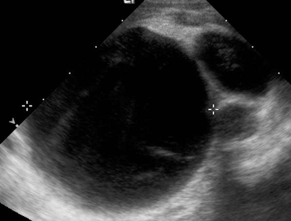
Ultrasonography: Multicystic dysplastic kidney
-
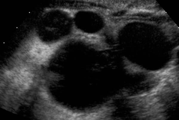
Ultrasonography: Multicystic dysplastic kidney

-
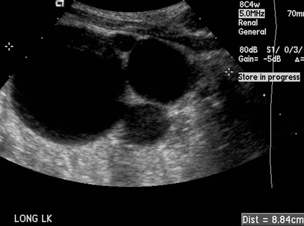
Ultrasonography: Multicystic dysplastic kidney
-
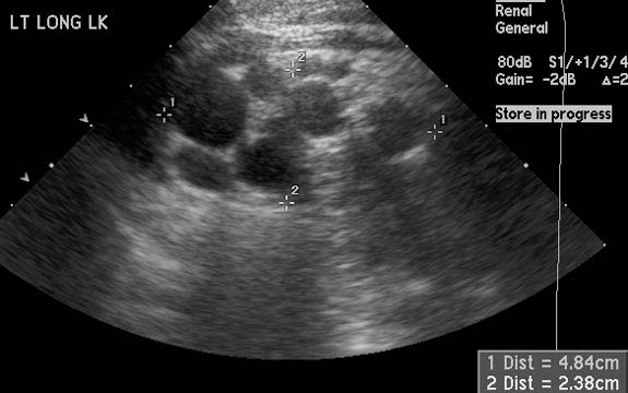
Ultrasonography: Multicystic dysplastic kidney


Treatment
MCDK is not treatable. However, the patient is observed periodically for the first few years during which ultrasounds are generally taken to ensure the healthy kidney is functioning properly and that the unhealthy kidney is not causing adverse effects.
External links
- Goldminer: Multicystic dysplastic kidney
- http://www.childrensmemorial.org/depts/fetalhealth/Fetal_MCDK.asp
- Template:Chorus
References
- ↑ Maria-Gisela Mercado-Deane, James E. Beeson, and Susan D. John. US of Renal Insufficiency in Neonates. RadioGraphics 2002 22: 1429-1438.
- ↑ http://www.childrensmemorial.org/depts/fetalhealth/Fetal_MCDK.asp