Medullary thyroid cancer biopsy: Difference between revisions
No edit summary |
No edit summary |
||
| Line 3: | Line 3: | ||
{{CMG}}; {{AE}} {{Ammu}} | {{CMG}}; {{AE}} {{Ammu}} | ||
==Overview== | ==Overview== | ||
On biopsy, medullary thyroid cancer is characterized by | On biopsy, medullary thyroid cancer is characterized by interstitial edema and coarse calcifications. | ||
==Key Biopsy Findings in Medullary Thyroid Cancer== | ==Key Biopsy Findings in Medullary Thyroid Cancer== | ||
===Microscopic Pathology=== | ===Microscopic Pathology=== | ||
| Line 10: | Line 9: | ||
====Cytoplasm and Nuclei==== | ====Cytoplasm and Nuclei==== | ||
:* Nested with delicate vascular septa | :* Nested with delicate vascular septa | ||
:* Trabecular | :* Trabecular cells | ||
:* Tubular/glandular, pseudo-papillary cells | :* Tubular/glandular, pseudo-papillary cells | ||
:* Polygonal to spindle to small cells | :* Polygonal to spindle to small cells | ||
:* Amphophilic, somewhat granular cytoplasm | :* Amphophilic, somewhat granular cytoplasm | ||
:* Interstitial edema | :* Interstitial edema | ||
:* | :* Vascular stroma may have amyloid deposits with fluffy appearing acellular eosinophilic material in the cytoplasm | ||
:* Stroma | :* Stroma can show hemorrhage, hyalinised collagen, oedema or metaplastic bone | ||
:* Coarse calcification and psammoma bodies may be present | :* Coarse calcification and psammoma bodies may be present | ||
:* Nuclei with "neuroendocrine features" | :* Nuclei with "neuroendocrine features" | ||
:* Small, round nuclei. | :* Small, round nuclei. | ||
:* Coarse chromatin (salt and pepper nuclei) | :* Coarse chromatin (salt and pepper nuclei) | ||
====Surrounding Thyroid==== | ====Surrounding Thyroid==== | ||
:* +/-C-cell hyperplasia - seen with familial forms of | :* +/-C-cell hyperplasia - seen with familial forms of medullary thyroid cancer | ||
:* C cells (AKA parafollicular cell): abundant cytoplasm - clear/pale | :* C cells (AKA parafollicular cell): abundant cytoplasm - clear/pale | ||
==Biopsy Exams of Medullary Thyroid Cancer== | ==Biopsy Exams of Medullary Thyroid Cancer== | ||
<gallery> | <gallery> | ||
| Line 62: | Line 61: | ||
Image:Thyroid MedullaryCarcinoma Amyloid HP PA.JPG| Medullary thyroid carcinoma<ref> Medullary thyroid cancer librepathology | Image:Thyroid MedullaryCarcinoma Amyloid HP PA.JPG| Medullary thyroid carcinoma<ref> Medullary thyroid cancer librepathology | ||
(2015).http://librepathology.org/wiki/index.php/Medullary_thyroid_carcinoma.JPG Accessed on November 12, 2015</ref> | (2015).http://librepathology.org/wiki/index.php/Medullary_thyroid_carcinoma.JPG Accessed on November 12, 2015</ref> | ||
</gallery> | </gallery> | ||
==References== | ==References== | ||
{{Reflist|2}} | {{Reflist|2}} | ||
Revision as of 19:29, 18 November 2015
|
Medullary thyroid cancer Microchapters |
|
Differentiating Medullary thyroid cancer from other Diseases |
|---|
|
Diagnosis |
|
Treatment |
|
Case Studies |
|
Medullary thyroid cancer biopsy On the Web |
|
American Roentgen Ray Society Images of Medullary thyroid cancer biopsy |
|
Risk calculators and risk factors for Medullary thyroid cancer biopsy |
Editor-In-Chief: C. Michael Gibson, M.S., M.D. [1]; Associate Editor(s)-in-Chief: Ammu Susheela, M.D. [2]
Overview
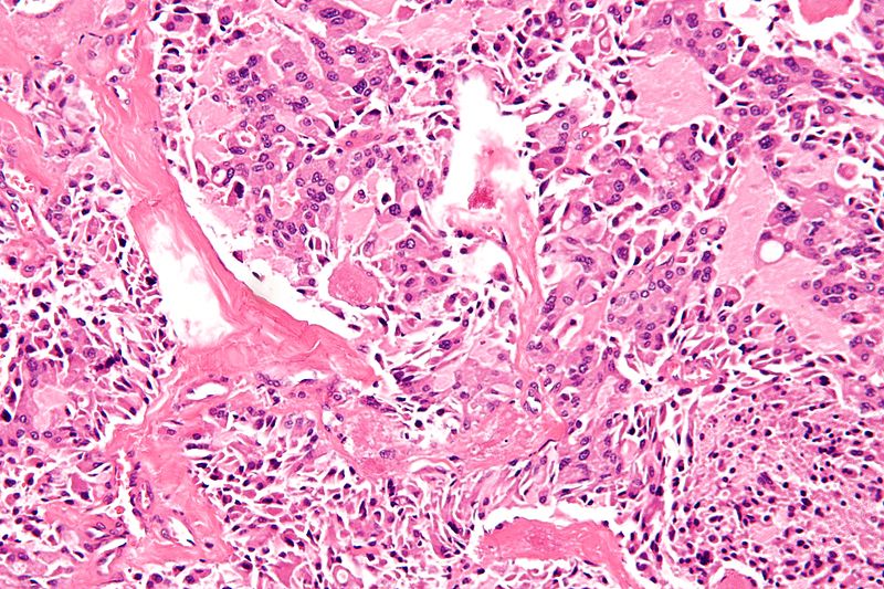
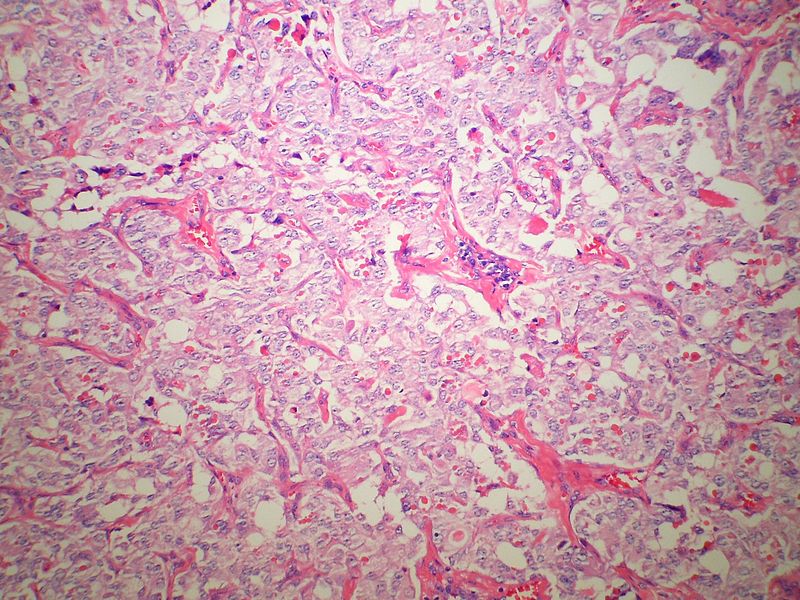
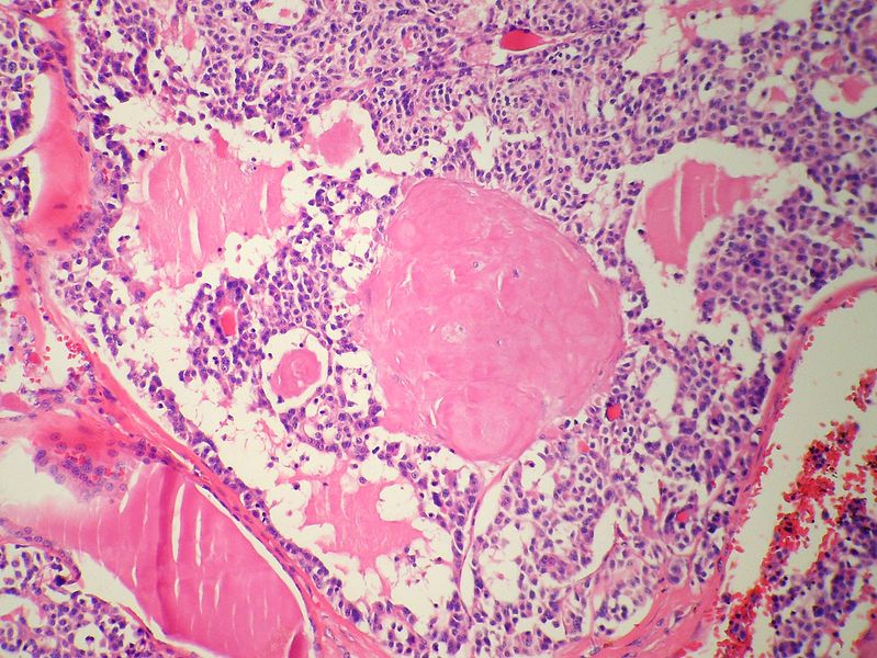
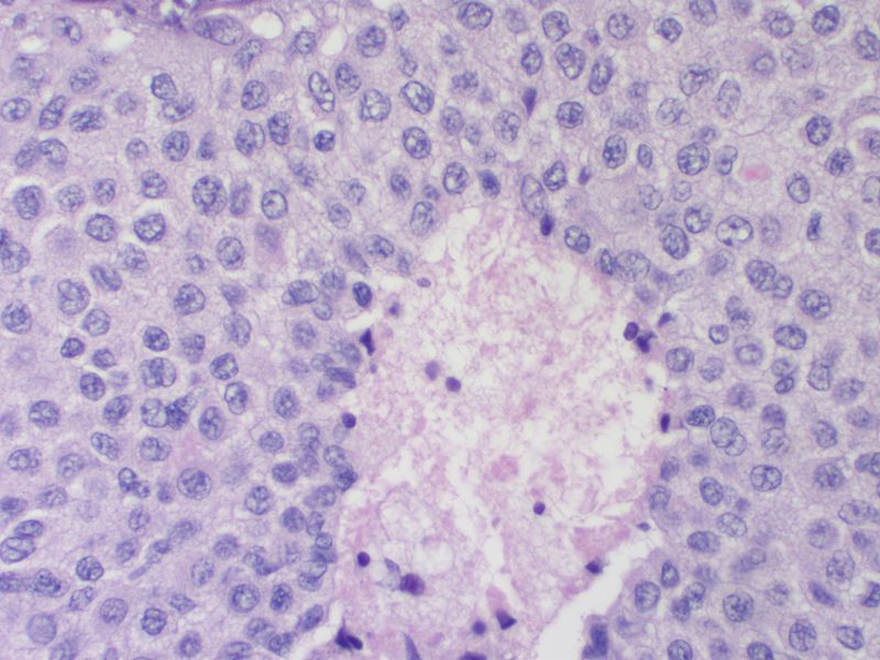
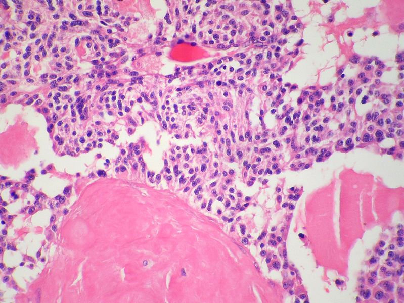
On biopsy, medullary thyroid cancer is characterized by interstitial edema and coarse calcifications.
Key Biopsy Findings in Medullary Thyroid Cancer
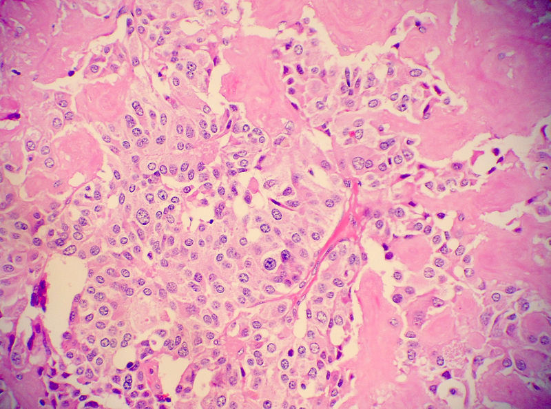
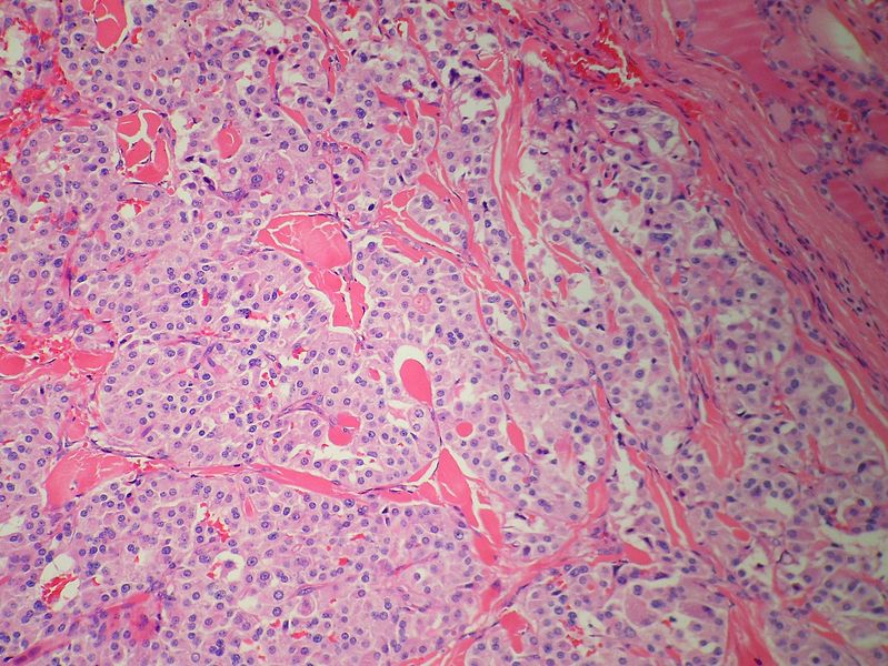
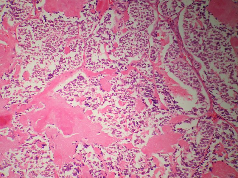
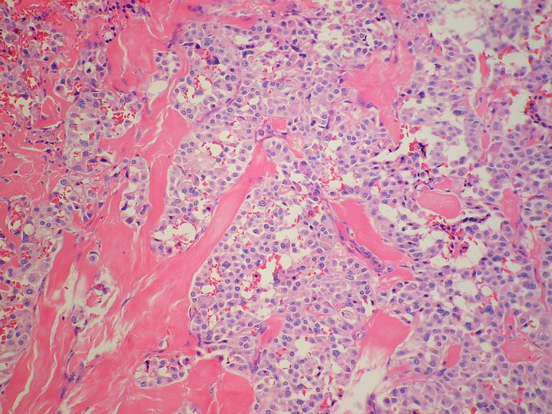
Microscopic Pathology
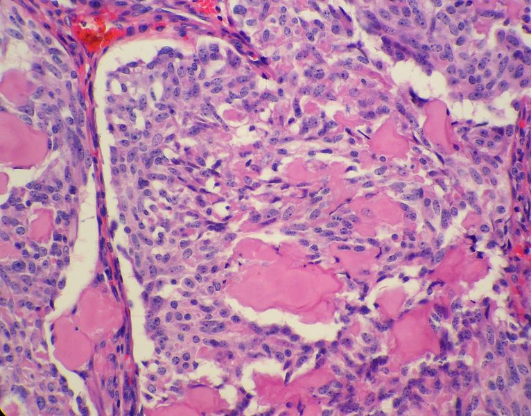
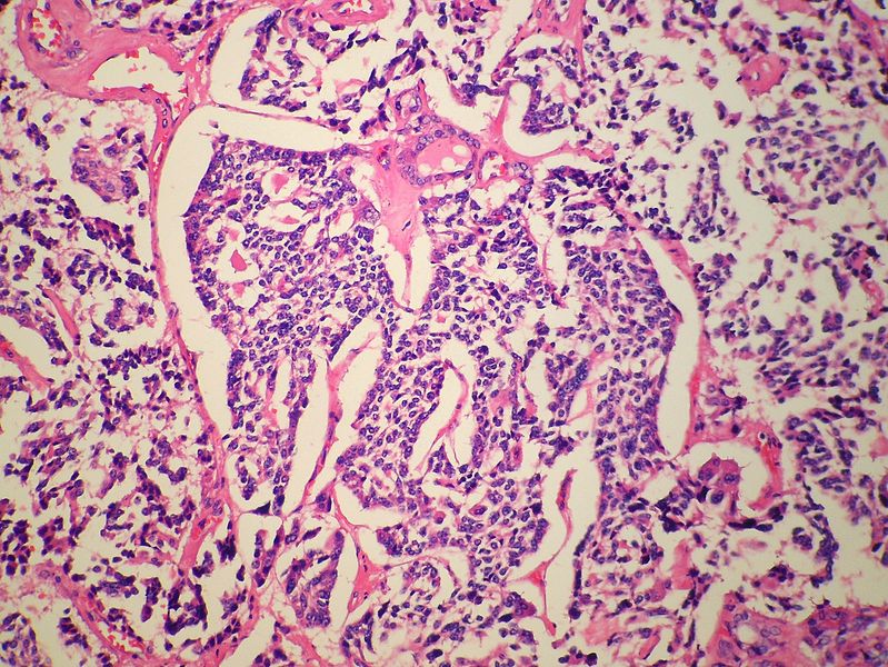
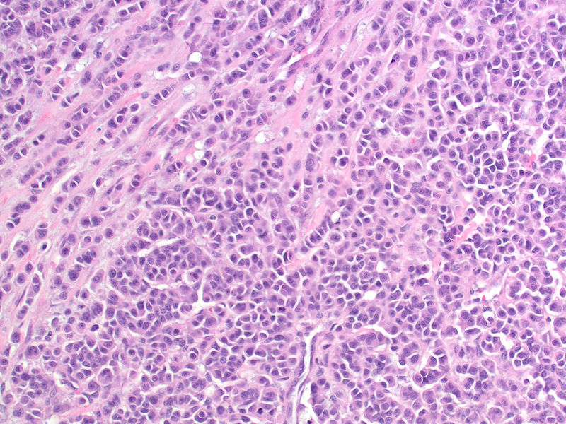
- Microscopic features of medullary thyroid cancer is as follows:
Cytoplasm and Nuclei
- Nested with delicate vascular septa
- Trabecular cells
- Tubular/glandular, pseudo-papillary cells
- Polygonal to spindle to small cells
- Amphophilic, somewhat granular cytoplasm
- Interstitial edema
- Vascular stroma may have amyloid deposits with fluffy appearing acellular eosinophilic material in the cytoplasm
- Stroma can show hemorrhage, hyalinised collagen, oedema or metaplastic bone
- Coarse calcification and psammoma bodies may be present
- Nuclei with "neuroendocrine features"
- Small, round nuclei.
- Coarse chromatin (salt and pepper nuclei)
Surrounding Thyroid
- +/-C-cell hyperplasia - seen with familial forms of medullary thyroid cancer
- C cells (AKA parafollicular cell): abundant cytoplasm - clear/pale
Biopsy Exams of Medullary Thyroid Cancer
-
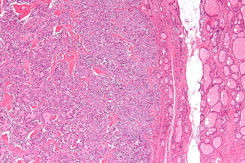
Low magnification micrograph of medullary thyroid carcinoma<ref> Medullary thyroid cancer librepathology
-
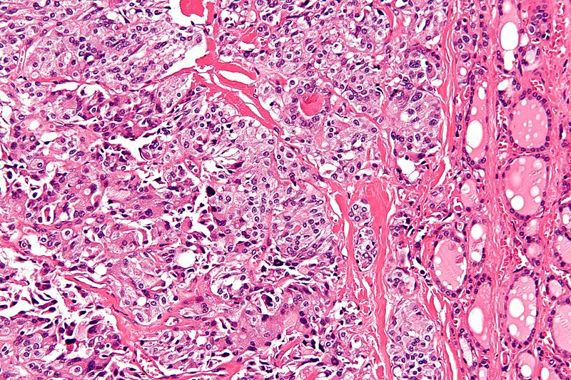
High magnification micrograph of medullary thyroid carcinoma<ref> Medullary thyroid cancer librepathology
-
High magnification micrograph of medullary thyroid carcinoma<ref> Medullary thyroid cancer librepathology
-
Low magnification micrograph of medullary thyroid carcinoma<ref> Medullary thyroid cancer librepathology
-
Medullary thyroid carcinoma amyloid<ref> Medullary thyroid cancer librepathology
-
Medullary thyroid carcinoma amyloid<ref> Medullary thyroid cancer librepathology
-
Medullary thyroid carcinoma amyloid<ref> Medullary thyroid cancer librepathology
-
Medullary thyroid carcinoma amyloid<ref> Medullary thyroid cancer librepathology
-
Medullary thyroid carcinoma amyloid<ref> Medullary thyroid cancer librepathology
-
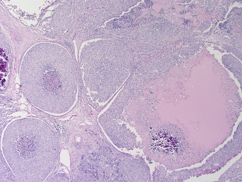
Medullary thyroid carcinoma comedonecrosis<ref> Medullary thyroid cancer librepathology
-
Medullary thyroid carcinoma comedonecrosis<ref> Medullary thyroid cancer librepathology
-

Medullary thyroid carcinoma comedonecrosis<ref> Medullary thyroid cancer librepathology
-
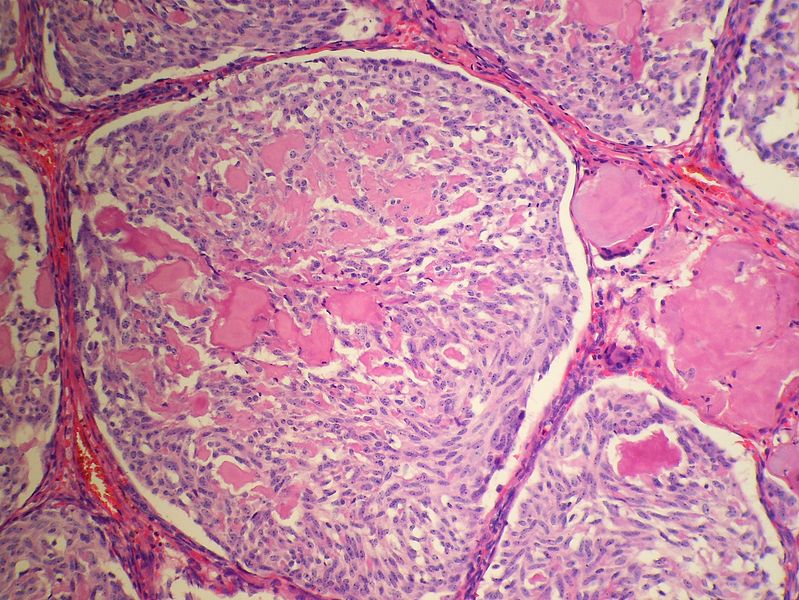
Medullary thyroid carcinoma spindle cell<ref> Medullary thyroid cancer librepathology
-
Medullary thyroid carcinoma spindle cell<ref> Medullary thyroid cancer librepathology
-
Medullary thyroid carcinoma spindle cell<ref> Medullary thyroid cancer librepathology
-
Medullary thyroid carcinoma spindle cell<ref> Medullary thyroid cancer librepathology
-
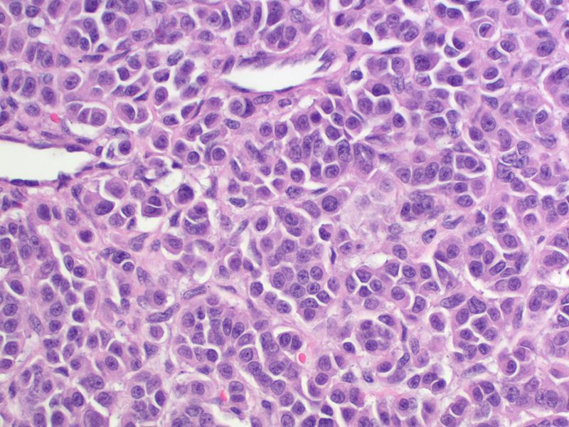
Medullary thyroid carcinoma spindle cell<ref> Medullary thyroid cancer librepathology
-
Medullary thyroid carcinoma<ref> Medullary thyroid cancer librepathology


.jpg)
