Liver mass CT: Difference between revisions
Aditya Ganti (talk | contribs) (→CT) |
Aditya Ganti (talk | contribs) (→CT) |
||
| Line 5: | Line 5: | ||
==Overview== | ==Overview== | ||
Computed tomography may be useful for the evaluation and diagnosis of liver masses. The evaluation of liver mass should be performed with a triphasic CT, this modality includes 3 phases: non-contrast, arterial phase, and portal venous phase. On CT, characteristic findings of liver mass, may include: solitary or multiple lesion, solid or cystic consistency, and normally a rounded lesion. The evaluation of liver mass will depend on several characteristics, such as: vascular pattern, size, location, size, distribution, margins, attenuation, and contrast enhancement. | Computed tomography may be useful for the evaluation and diagnosis of liver masses. The evaluation of liver mass should be performed with a triphasic CT, this modality includes 3 phases: non-contrast, arterial phase, and portal venous phase. On CT, characteristic findings of liver mass, may include: solitary or multiple lesion, solid or cystic consistency, and normally a rounded lesion. The evaluation of liver mass will depend on several characteristics, such as: vascular pattern, size, location, size, distribution, margins, attenuation, and contrast enhancement. | ||
==CT== | ==CT== | ||
Computed tomography may be useful for the evaluation and diagnosis of liver masses. | Computed tomography may be useful for the evaluation and diagnosis of liver masses.<ref name="radioas" /><ref name="pmid22541698">{{cite journal |vauthors=Bonder A, Afdhal N |title=Evaluation of liver lesions |journal=Clin Liver Dis |volume=16 |issue=2 |pages=271–83 |year=2012 |pmid=22541698 |doi=10.1016/j.cld.2012.03.001 |url=}}</ref> | ||
*The evaluation of liver mass should be performed with a triphasic CT, this modality includes 3 phases: | *The evaluation of liver mass should be performed with a triphasic CT, this modality includes 3 phases: | ||
**Non-contrast | **Non-contrast | ||
| Line 18: | Line 18: | ||
**Solid or cystic | **Solid or cystic | ||
**Rounded lesion | **Rounded lesion | ||
**'''Bright dot sign''': Presence of a bright dot within a lesion which remains hyper-attenuating on arterial and portal venous phase CT, corresponding to early nodular enhancement seen on liver hemangioma. | |||
{| class="wikitable" | {| class="wikitable" | ||
! | !Common Liver masses | ||
!CT scan Findings | !CT scan Findings | ||
|- | |- | ||
|[[Hepatocellular carcinoma]] | |||
| | |||
*Early arterial phase enhancement and then rapid wash out | *Early arterial phase enhancement and then rapid wash out | ||
*Rim enhancement of capsule may persist | *Rim enhancement of capsule may persist | ||
*Malignant liver mass, particularly hepatocellular carcinoma, can have a variety of appearances, such as: | |||
**Massive (focal) | |||
*Large mass | ***Large mass | ||
*May have necrosis, fat and /or calcification | ***May have necrosis, fat and /or calcification | ||
**Nodular (multifocal) | |||
***Multiple masses of variable attenuation | |||
***May also have central necrosis | |||
**Infiltrative (diffuse) | |||
|- | |- | ||
|[[Hemangioma]] | |||
| | | | ||
*Discontinuous, nodular, peripheral enhancement starting in arterial phase | *Discontinuous, nodular, peripheral enhancement starting in arterial phase | ||
| Line 46: | Line 43: | ||
*Small hemangiomas (< ~1.5 cm) may demonstrate "flash filling" - complete homogenous enhancement in arterial phase (no gradual filling in) | *Small hemangiomas (< ~1.5 cm) may demonstrate "flash filling" - complete homogenous enhancement in arterial phase (no gradual filling in) | ||
|- | |- | ||
|[[Focal nodular hyperplasia]] | |||
| | | | ||
*Bright arterial phase enhancement except central scar | *Bright arterial phase enhancement except central scar | ||
| Line 52: | Line 49: | ||
*Central scar enhancement on delayed phase | *Central scar enhancement on delayed phase | ||
|- | |- | ||
|[[Hepatic adenoma]] | |||
| | | | ||
*Large, well circumscribed encapsulated tumors | *Large, well circumscribed encapsulated tumors | ||
| Line 62: | Line 59: | ||
*Returns to near isodensity on portal venous and delayed phase image | *Returns to near isodensity on portal venous and delayed phase image | ||
|- | |- | ||
|[[Metastases|Liver metastases]] | |||
| | | | ||
*Hypodense and enhance less than the surrounding liver | *Hypodense and enhance less than the surrounding liver | ||
| Line 68: | Line 65: | ||
*Rim enhancement is a feature of malignant lesions, especially metastases. | *Rim enhancement is a feature of malignant lesions, especially metastases. | ||
|} | |} | ||
==Gallery== | ==Gallery== | ||
Revision as of 20:25, 29 January 2018
|
Liver Mass Microchapters |
|
Diagnosis |
|---|
|
Treatment |
|
Case Studies |
|
Liver mass CT On the Web |
|
American Roentgen Ray Society Images of Liver mass CT |
Editor-In-Chief: C. Michael Gibson, M.S., M.D. [1]Associate Editor(s)-in-Chief: Maria Fernanda Villarreal, M.D. [2]
Overview
Computed tomography may be useful for the evaluation and diagnosis of liver masses. The evaluation of liver mass should be performed with a triphasic CT, this modality includes 3 phases: non-contrast, arterial phase, and portal venous phase. On CT, characteristic findings of liver mass, may include: solitary or multiple lesion, solid or cystic consistency, and normally a rounded lesion. The evaluation of liver mass will depend on several characteristics, such as: vascular pattern, size, location, size, distribution, margins, attenuation, and contrast enhancement.
CT
Computed tomography may be useful for the evaluation and diagnosis of liver masses.[1][2]
- The evaluation of liver mass should be performed with a triphasic CT, this modality includes 3 phases:
- Non-contrast
- Arterial phase
- Portal venous phase
- On CT, characteristic findings of liver mass, include:[1]
- Solitary or multiple lesion
- Solid or cystic
- Rounded lesion
- Bright dot sign: Presence of a bright dot within a lesion which remains hyper-attenuating on arterial and portal venous phase CT, corresponding to early nodular enhancement seen on liver hemangioma.
| Common Liver masses | CT scan Findings |
|---|---|
| Hepatocellular carcinoma |
|
| Hemangioma |
|
| Focal nodular hyperplasia |
|
| Hepatic adenoma |
|
| Liver metastases |
|
Gallery
-
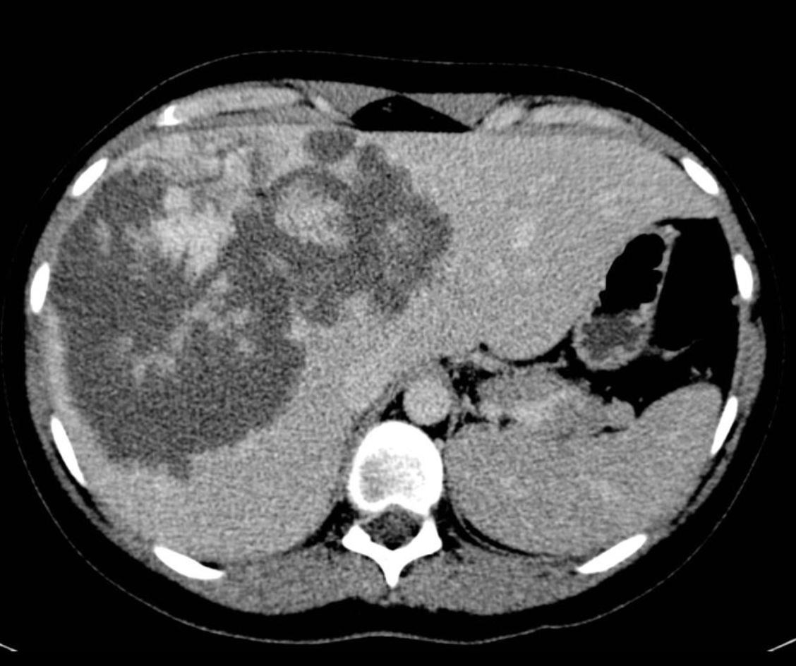
Liver hemangioma: discontinuous, nodular, peripheral enhancement starting in arterial phase
-
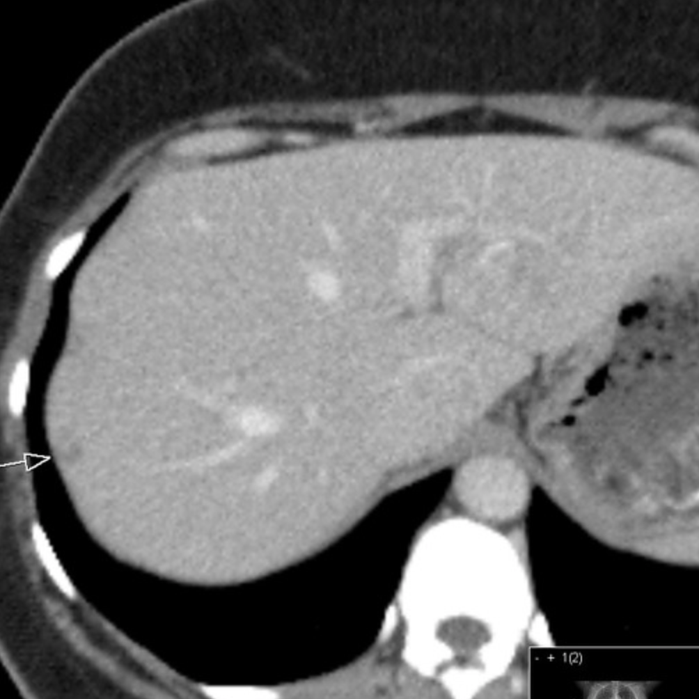
Bright dot sign: Bright dot within a lesion which remains hyper-attenuating on arterial and portal venous phase CT, corresponding to early nodular enhancement seen on liver hemangioma
-
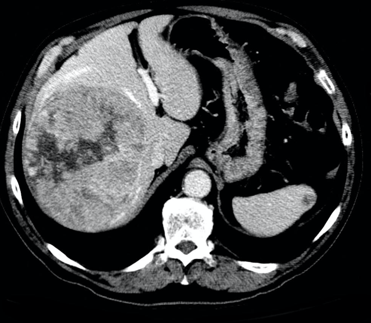
Focal nodular hyperplasia: bright arterial contrast enhancement except for the central scar which remains hypoattenuating
-

Hepatic abscess: peripheral enhancement, centrally hypoattenuating lesions. Occasionally they appear solid, or contain gas. Segmental perfusion abnormalities, with early enhancement, may be seen.


References
- ↑ 1.0 1.1 Oliver JH, Baron RL: State of the art, helical biphasic contrast enhanced CT of the liver: Technique, indications, interpretation, and pitfalls. Radiology 1996; 201:1-14.
- ↑ Bonder A, Afdhal N (2012). "Evaluation of liver lesions". Clin Liver Dis. 16 (2): 271–83. doi:10.1016/j.cld.2012.03.001. PMID 22541698.