Listeria monocytogenes: Difference between revisions
Joao Silva (talk | contribs) |
Joao Silva (talk | contribs) |
||
| Line 39: | Line 39: | ||
*[[vomiting]] | *[[vomiting]] | ||
*headache | *headache | ||
*[[arthralgia|joint]] and muscle pain | *[[arthralgia|joint]] and [[myalgia|muscle pain]] | ||
It generally lasts for 2 days and the patient experiences complete recovery from the symptoms. Some patients may be asymptomatic for the disease, whilst others, immunocompromised, pregnant women and elder patients, in rare cases, present with invasive infection. | It generally lasts for 2 days and the patient experiences complete recovery from the symptoms. Some patients may be asymptomatic for the disease, whilst others, immunocompromised, pregnant women and elder patients, in rare cases, present with invasive infection. | ||
Revision as of 20:03, 23 February 2014
| Listeria monocytogenes | ||||||||||||||
|---|---|---|---|---|---|---|---|---|---|---|---|---|---|---|
 | ||||||||||||||
| Scientific classification | ||||||||||||||
| ||||||||||||||
| Binomial name | ||||||||||||||
| Listeria monocytogenes Murray et al. (1926) |
Editor-In-Chief: C. Michael Gibson, M.S., M.D. [1]; Associate Editor(s)-in-Chief: João André Alves Silva, M.D. [2]
Overview
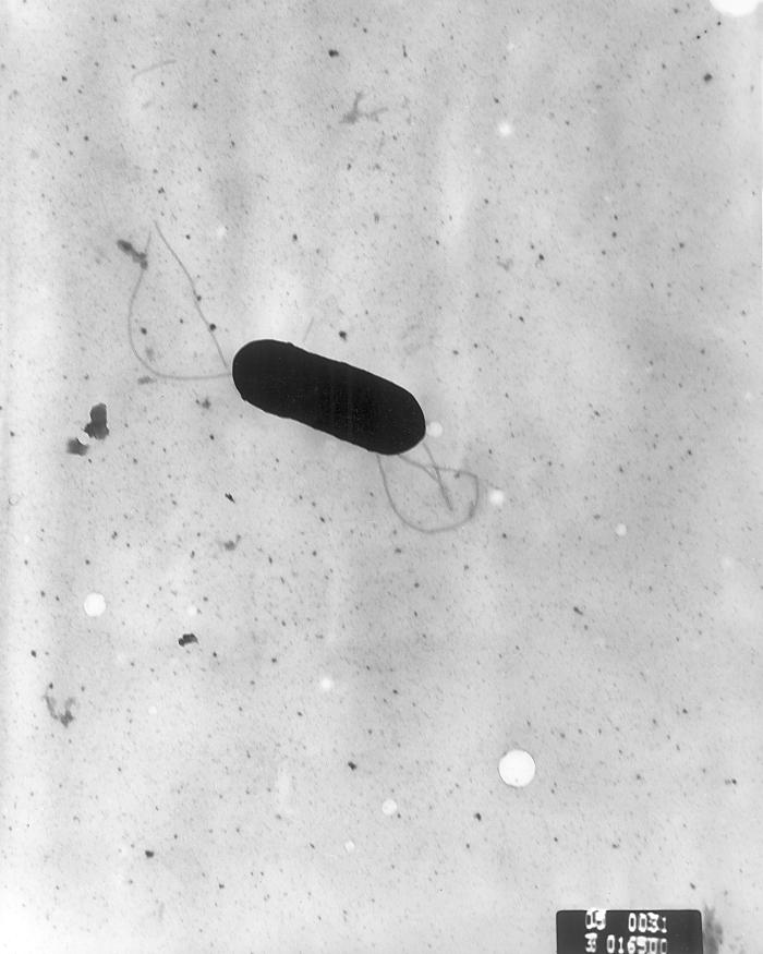
Listeria monocytogenes is a Gram-positive bacterium, in the division Firmicutes, named for Joseph Lister. Motile via flagella, L. monocytogenes can move within eukaryotic cells by explosive polymerization of actin filaments (known as comet tails or actin rockets).
Studies suggest that up to 10% of human gastrointestinal tracts may be colonized by L. monocytogenes.
Clinical Manifestations
Listeria monocytogenes is known for causing disease particularly in immunosuppressed patients, neonates, elderly, pregnant women and occasionally in healthy individuals. It has an incubation period which can range from a median of 24 hours, in Listeria gastroenteritis, to a median of 35 days, in Listeria invasive disease. The infection with Listeria monocytogenes is more common to occur during the summer and may present as a self-limited febrile gastroenteritis, in normal adult hosts, after ingesting a high number of organisms in food, or as an invasive disease.
Febrile Gastroenteritis
Febrile gastroenteritis accounts for less than 1% of reported bacterial food-born infections, occurring usually after ingestion of a large inoculum of bacteria from contaminated foods. It might present as:
- fever
- watery diarrhea
- nausea
- vomiting
- headache
- joint and muscle pain
It generally lasts for 2 days and the patient experiences complete recovery from the symptoms. Some patients may be asymptomatic for the disease, whilst others, immunocompromised, pregnant women and elder patients, in rare cases, present with invasive infection.
Infection in Pregnancy
Occurs more frequently during the third trimester of gestation, presenting most commonly with flu-like symptoms, such as:
- fever
- chills
- back pain
The infection may be mild and the diagnosis missed, if blood cultures are not obtained. Since bacteremia, with no CNS involvement is common rule in pregnant women with listeriosis, blood cultures should always be obtained in pregnant women who present with fever, with no other alternative cause, such as UTI or pharyngitis.
Listeriosis, in pregnant women, can result in:
- fetal death
- premature birth
- infected newborns
The newborn also has great risk of developing granulomatosis infantiseptica, a severe 'in utero' infection resulting from transplacental transmission, in which infants may present with:
- disseminated abscesses
- granulomas in multiple internal organs (brain, lungs, liver, spleen and kidneys)
- papular or ulcerative skin lesions.
- most infants with this disease are stillborn or die soon after birth.
Sepsis of Unknown Origin
CNS Infection
- Meningoencephalitis
- Cerebritis
- Rhombencephalitis
Focal infections
Diagnosis
Pathogenesis
Infection by L. monocytogenes causes the disease listeriosis. The manifestations of listeriosis include septicemia[1], meningitis (or meningoencephalitis)[1], encephalitis[2], corneal ulcer[3], Pneumonia[4], and intrauterine or cervical infections in pregnant women, which may result in spontaneous abortion (2nd/3rd trimester) or stillbirth. Surviving neonates of Fetomaternal Listeriosis may suffer granulomatosis infantiseptica - pyogenic granulomas distributed over the whole body, and may suffer from physical retardation. Influenza-like symptoms, including persistent fever, usually precede the onset of the aforementioned disorders. Gastrointestinal symptoms such as nausea, vomiting, and diarrhea may precede more serious forms of listeriosis or may be the only symptoms expressed. Gastrointestinal symptoms were epidemiologically associated with use of antacids or cimetidine. The onset time to serious forms of listeriosis is unknown but may range from a few days to three weeks. The onset time to gastrointestinal symptoms is unknown but probably exceeds 12 hours. An early study suggeseted that L. monocytogenes was unique among Gram-positive bacteria in that it possessed lipopolysaccharide[5], which served as an endotoxin. A later study did not support these findings[6].
The infective dose of L. monocytogenes varies with the strain and with the susceptibility of the victim. From cases contracted through raw or supposedly pasteurized milk, one may safely assume that in susceptible persons, fewer than 1,000 total organisms may cause disease. L. monocytogenes may invade the gastrointestinal epithelium. Once the bacterium enters the host's monocytes, macrophages, or polymorphonuclear leukocytes, it becomes blood-borne (septicemic) and can grow. Its presence intracellularly in phagocytic cells also permits access to the brain and probably transplacental migration to the fetus in pregnant women. The pathogenesis of L. monocytogenes centers on its ability to survive and multiply in phagocytic host cells.
Treatment
When listeric meningitis occurs, the overall mortality may reach 70%; from septicemia 50%, from perinatal/neonatal infections greater than 80%. In infections during pregnancy, the mother usually survives. Reports of successful treatment with parenteral penicillin or ampicillin exist. Trimethoprim-sulfamethoxazole has been shown effective in patients allergic to penicillin.
Bacteriophage treatments have been developed by several companies. EBI Food Safety and Intralytix both have products suitable for treatment of the bacteria. The FDA of the United States approved a cocktail of six bacteriophages from Intralytix, and a one type phage product from EBI Food Safety designed to kill the bacteria L. monocytogenes. Uses would potentially include spraying it on fruits and ready-to-eat meat such as sliced ham and turkey.
Gene Therapy
L. monocytogenes has been used in studies to deliver genes in vitro. However transfection efficiency remains poor.
Detection

The methods for analysis of food are complex and time-consuming. The present Food and Drug Administration (FDA) method, revised in September, 1990, requires 24 and 48 hours of enrichment, followed by a variety of other tests. Total time to identification takes from 5 to 7 days, but the announcement of specific nonradiolabled DNA probes should soon allow a simpler and faster confirmation of suspect isolates.
Recombinant DNA technology may even permit 2-to-3 day positive analysis in the future. Currently, the FDA is collaborating in adapting its methodology to quantitate very low numbers of the organisms in foods.
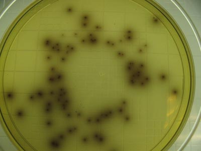
Bio-Rad Laboratories (www.bio-rad.com) have come up with media called Rapid'L.Mono Medium which cut short time to 48 hours
Epidemiology
Researchers have found L. monocytogenes in at least 37 mammalian species, both domesticated and feral, as well as in at least 17 species of birds and possibly in some species of fish and shellfish. Laboratories can isolate L. monocytogenes from soil, silage, and other environmental sources. L. monocytogenes is quite hardy and resists the deleterious effects of freezing, drying, and heat remarkably well for a bacterium that does not form spores. Most L. monocytogenes are pathogenic to some degree.
Routes of infection
L. monocytogenes has been associated with such foods as raw milk, pasteurized fluid milk[7], cheeses (particularly soft-ripened varieties), ice cream, raw vegetables, fermented raw-meat sausages, raw and cooked poultry, raw meats (of all types), and raw and smoked fish. Its ability to grow at temperatures as low as 0°C permits multiplication in refrigerated foods. In refrigeration temperature such as 4°C the amount of ferric iron promotes the growth of L. monocytogenes.[8]
Infectious Cycle
The primary site of infection is the intestinal epithelium where the bacteria invade non-phagocytic cells via the "zipper" mechanism. Uptake is stimulated by the binding of listerial internalins (Inl) to host cell adhesion factors such as E-cadherin or Met. This binding activates certain Rho-GTPases which subsequently bind and stabilize Wiskott Aldrich Syndrome Protein (WASp). WASp can then bind the Arp2/3 complex and serve as an actin nucleation point. Subsequent actin polymerization extends the cell membrane around the bacterium, eventually engulfing it. The net effect of internalin binding is to exploit the junction forming-apparatus of the host into internalizing the bacterium. Note that L. monocytogenes can also invade phagocytic cells (e.g. macrophages) but only requires internalins for invasion of non-phagocytic cells.
Following internalisation, the bacterium must escape from the vacuole/phagosome before fusion with a lysosome can occur. Two main virulence factors which allow the bacterium to escape are listeriolysin O (LLO - encoded by hly) and phospholipase C B (plcB). Secretion of LLO and PlcB disrupts the vacuolar membrane and allows the bacterium to escape into the cytoplasm where it may proliferate.
Once in the cytoplasm, L. monocytogenes exploits host actin for the second time. ActA proteins associated with the old bacterial cell pole (being a bacilli, L. monocytogenes septates in the middle of the cell and thus has one new pole and one old pole) are capable of binding the Arp2/3 complex and thus induce actin nucleation at a specific area of the bacterial cell surface. Actin polymerization then propels the bacterium unidirectionally into the host cell membrane. The protrusion which is formed may then be internalised by a neighbouring cell, forming a double-membrane vacuole from which the bacterium must escape using LLO and PlcB.
References
- ↑ 1.0 1.1 Gray, M. L., and A. H. Killinger. 1966. Listeria monocytogenes and listeric infection. Bacteriol. Rev. 30:309-382.
- ↑ Armstrong, R. W., and P. C. Fung. 1993. Brainstem encephalitis (Rhombencephalitis) due to Listeria monocytogenes: case report and review. Clin. Infect. Dis. 16:689-702.
- ↑ Holland, S., E. Alfonso, H. Gelender, D. Heidemann, A. Mendelsohn, S. Ullman, and D. Miller. 1987. Corneal ulcer due to Listeria monocytogenes. Cornea 6:144-146.
- ↑ Whitelock-Jones, L., J. Carswell, and K. C. Rassmussen. 1989. Listeria pneumonia. A case report. South African Medical Journal 75:188-189.
- ↑ Wexler, H., and J. D. Oppenheim. 1979. Isolation, characterization, and biological properties of an endotoxin-like material from the gram-positive organism Listeria monocytogenes. Infect. Immun. 23:845-857.
- ↑ SHYAMAL K. MAITRA,RONALD NACHUM, AND FREDERICK C. PEARSON. 1986. Establishment of Beta-Hydroxy Fatty Acids as Chemical Marker Molecules for Bacterial Endotoxin by Gas Chromatography-Mass Spectrometry. APPLIED AND ENVIRONMENTAL MICROBIOLOGY, Sept. 1986, p. 510-514
- ↑ Fleming, D. W., S. L. Cochi, K. L. MacDonald, J. Brondum, P. S. Hayes, B. D. Plikaytis, M. B. Holmes, A. Audurier, C. V. Broome, and A. L. Reingold. 1985. Pasteurized milk as a vehicle of infection in an outbreak of listeriosis. N. Engl. J. Med. 312:404-407.
- ↑ Dykes, G. A., Dworaczek (Kubo), M. 2002. Influence of interactions between temperature, ferric ammonium citrate and glycine betaine on the growth of Listeria monocytogenes in a defined medium. Lett Appl Microbiol. 35(6):538-42.
External links
cs:Listeria monocytogenes de:Listeria monocytogenes ko:리스테리아 nl:Listeria monocytogenes