Listeria monocytogenes: Difference between revisions
YazanDaaboul (talk | contribs) |
YazanDaaboul (talk | contribs) (→Cause) |
||
| Line 34: | Line 34: | ||
==Cause== | ==Cause== | ||
*Listeriosis is caused by the bacterium ''Listeria spp | *Listeriosis is caused by the bacterium ''Listeria spp''. | ||
*''Listeria monocytogenes'' is the most common species associated with development of listeriosis. | *''Listeria monocytogenes'' is the most common species associated with development of listeriosis. | ||
*The genus ''Listeria'' contains ten species: | *The genus ''Listeria'' contains ten species: | ||
Latest revision as of 17:11, 25 January 2016
|
Listeriosis Microchapters |
|
Diagnosis |
|---|
|
Treatment |
|
Case Studies |
|
Listeria monocytogenes On the Web |
|
American Roentgen Ray Society Images of Listeria monocytogenes |
|
Risk calculators and risk factors for Listeria monocytogenes |
| Listeria | ||||||||||||
|---|---|---|---|---|---|---|---|---|---|---|---|---|
 Scanning electron micrograph of Listeria monocytogenes.
| ||||||||||||
| Scientific classification | ||||||||||||
| ||||||||||||
| Species | ||||||||||||
|
L. fleischmannii |
Editor-In-Chief: C. Michael Gibson, M.S., M.D. [1]; Associate Editor(s)-in-Chief: João André Alves Silva, M.D. [2]
Overview
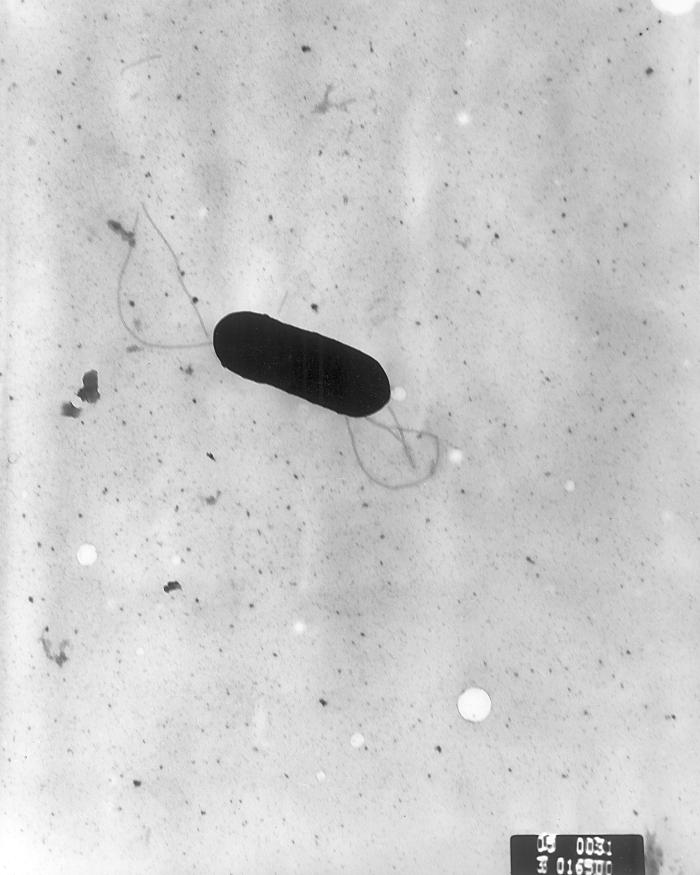
Listeriosis is caused by the bacterium Listeria monocytogenes, a flagellated, catalase-positive, facultative intracellular, anaerobic, nonsporulating, Gram-positive bacillus. Listeria is commonly found in soil, water, vegetation and fecal material.[1]
Cause
- Listeriosis is caused by the bacterium Listeria spp.
- Listeria monocytogenes is the most common species associated with development of listeriosis.
- The genus Listeria contains ten species:
- L. fleischmannii
- L. grayi
- L. innocua
- L. ivanovii
- L. marthii
- L. monocytogenes
- L. rocourtiae
- L. seeligeri
- L. weihenstephanensis
- L. welshimeri
- Of note, Listeria dinitrificans was previously thought to be part of the Listeria genus, but it has been reclassified into the new genus Jonesia.[2]
Taxonomy
Bacteria; Firmicutes; Bacilli; Bacillales; Listeriaceae; Listeria; Listeria monocytogenes.
Microbiological Characteristics
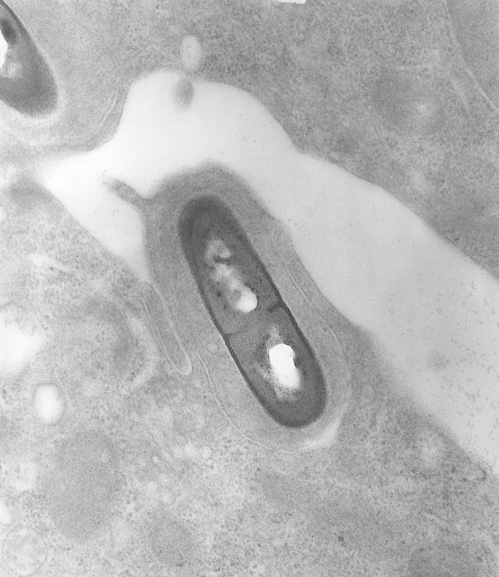
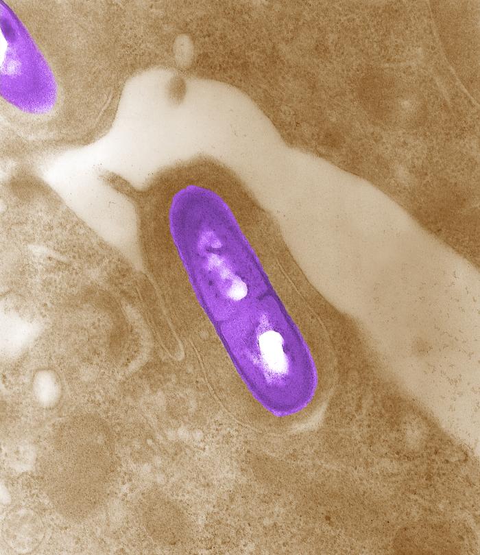
- Listeria monocytogenes is a flagellated, catalase-positive, facultative intracellular, anaerobic, nonsporulating, Gram-positive bacillus.
Natural Reservoir
- In the environment, Listeria monocytogenes is commonly found in soil, water, vegetation and fecal material.
- Animals may be asymptomatic carriers of Listeria.[1]
- L. monocytogenes has been associated with foods such as raw milk, pasteurized fluid milk, cheeses (particularly soft-ripened varieties), ice cream, raw vegetables, fermented raw-meat sausages, raw and cooked poultry, raw meats, and raw and smoked fish.[3]
- Listeria has the ability to grow at temperatures as low as 0°C, allows its multiplication in refrigerated foods. At refrigerated temperature such as 4°C, the amount of ferric iron in the environment promotes the growth of L. monocytogenes.[4]
Gallery


References
- ↑ 1.0 1.1 "Risk assessment of Listeria monocytogenes in ready-to-eat foods" (PDF).
- ↑ M. D. Collins, S. Wallbanks, D. J. Lane, J. Shah, R. Nietupskin, J. Smida, M. Dorsch and E. Stackebrandt. Phylogenetic Analysis of the Genus Listeria Based on Reverse Transcriptase Sequencing of 16S rRNA. International Journal of Systematic and Evolutionary Microbiology. April 1991 vol. 41 no. 2 240–246
- ↑ Fleming, D. W., S. L. Cochi, K. L. MacDonald, J. Brondum, P. S. Hayes, B. D. Plikaytis, M. B. Holmes, A. Audurier, C. V. Broome, and A. L. Reingold. 1985. Pasteurized milk as a vehicle of infection in an outbreak of listeriosis. N. Engl. J. Med. 312:404-407.
- ↑ Dykes, G. A., Dworaczek (Kubo), M. 2002. Influence of interactions between temperature, ferric ammonium citrate and glycine betaine on the growth of Listeria monocytogenes in a defined medium. Lett Appl Microbiol. 35(6):538-42.
- ↑ 5.0 5.1 "Public Health Image Library (PHIL), Centers for Disease Control and Prevention".