Left ventricular aneurysm CT: Difference between revisions
No edit summary |
No edit summary |
||
| Line 1: | Line 1: | ||
__NOTOC__ | __NOTOC__ | ||
{{Left ventricular aneurysm}} | {{Left ventricular aneurysm}} | ||
{{CMG}} | {{CMG}};{{AE}}{{MehdiP}} | ||
==Overview== | |||
Chest CT scan with or without contrast may reveal the size and the location of LV aneurysm also, it can show the presence of calcification on it. | |||
==CT== | ==CT== | ||
CT scan may be helpful in the diagnosis of LV aneurysm. CT scan findings are suggestive for location, size and presence of thrombosis in aneurysm wall. | |||
<gallery> | <gallery> | ||
Image:Calcified-ventricular-aneurysm-002.jpg|Calcified left ventricular aneurysm | Image:Calcified-ventricular-aneurysm-002.jpg|Calcified left ventricular aneurysm | ||
Revision as of 16:01, 8 November 2016
|
Left ventricular aneurysm Microchapters |
|
Differentiating Left ventricular aneurysm from other Diseases |
|---|
|
Diagnosis |
|
Treatment |
|
Case Studies |
|
Left ventricular aneurysm CT On the Web |
|
American Roentgen Ray Society Images of Left ventricular aneurysm CT |
|
Risk calculators and risk factors for Left ventricular aneurysm CT |
Editor-In-Chief: C. Michael Gibson, M.S., M.D. [1];Associate Editor(s)-in-Chief: Seyedmahdi Pahlavani, M.D. [2]
Overview
Chest CT scan with or without contrast may reveal the size and the location of LV aneurysm also, it can show the presence of calcification on it.
CT
CT scan may be helpful in the diagnosis of LV aneurysm. CT scan findings are suggestive for location, size and presence of thrombosis in aneurysm wall.
-
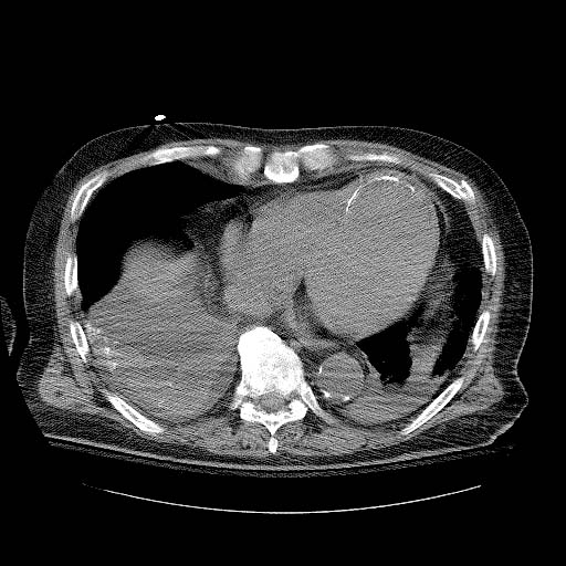
Calcified left ventricular aneurysm
-
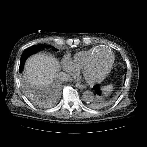
Calcified left ventricular aneurysm
-
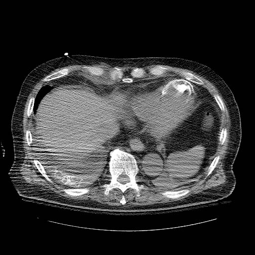
Calcified left ventricular aneurysm


