Keratoacanthoma
|
WikiDoc Resources for Keratoacanthoma |
|
Articles |
|---|
|
Most recent articles on Keratoacanthoma Most cited articles on Keratoacanthoma |
|
Media |
|
Powerpoint slides on Keratoacanthoma |
|
Evidence Based Medicine |
|
Clinical Trials |
|
Ongoing Trials on Keratoacanthoma at Clinical Trials.gov Trial results on Keratoacanthoma Clinical Trials on Keratoacanthoma at Google
|
|
Guidelines / Policies / Govt |
|
US National Guidelines Clearinghouse on Keratoacanthoma NICE Guidance on Keratoacanthoma
|
|
Books |
|
News |
|
Commentary |
|
Definitions |
|
Patient Resources / Community |
|
Patient resources on Keratoacanthoma Discussion groups on Keratoacanthoma Patient Handouts on Keratoacanthoma Directions to Hospitals Treating Keratoacanthoma Risk calculators and risk factors for Keratoacanthoma
|
|
Healthcare Provider Resources |
|
Causes & Risk Factors for Keratoacanthoma |
|
Continuing Medical Education (CME) |
|
International |
|
|
|
Business |
|
Experimental / Informatics |
Editor-In-Chief: C. Michael Gibson, M.S., M.D. [1];Associate Editor(s)-in-Chief: Kiran Singh, M.D. [2]
Overview
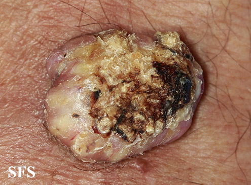
Keratoacanthoma (KA) is a relatively common, benign, epithelial tumor that was previously considered to be a variant of squamous cell carcinoma (SCC). The etiology is unknown. No human papillomavirus-DNA sequences were detected in lesions by polymerase chain reaction. It is a disease of the elderly (mean age, 64 years) with an annual incidence rate of 104 per 100,000. It is not associated with internal malignancy. There may be a seasonal presentation of keratoacanthoma that suggests that ultraviolet radiation has an acute effect on the development of KA. KAs may develop in sites of previous trauma. Most cases are the “crateriform” type, which grow rapidly then undergo spontaneous regression. Less than 2% belong to the rare destructive variants with no regression and persistent invasive growth. These are referred to as keratoacanthoma marginatum centrifugum and mutilating keratoacanthomas and can lead to severe defects.
KA begins as a smooth, dome-shaped, red papule that resembles molluscum contagiosum. In a few weeks the tumor may rapidly expand to 1 or 2cm and develop a central keratin-filled crater that is frequently filled with crust. The growth retains its smooth surface, unlike a squamous cell carcinoma. Untreated, growth stops in approximately 6 weeks, and the tumor remains unchanged for an indefinite period. In the majority of cases it then regresses slowly over 2 to 12 months and frequently heals with scarring. The limbs, particularly the sun-exposed hands and arms, are the most common site; the trunk is the second most common site, but KA may occur on any skin surface, including the anal area. On occasion, multiple KAs appear, or a single lesion extends over several centimeters. These variants resist treatment and are unlikely to undergo spontaneous emission.
According to a review of literature by Robert A. Schwartz, KA was once considered a benign neoplasm that resembled a highly malignant one (pseudomalignancy), but it is now viewed in an opposite light as a cancer that resembles a benign neoplasm (pseudobenignity). KA is an abortive malignancy that rarely progresses into an invasive SCC. The KA may serve as a marker for the important autosomal dominant familial cancer syndrome, the Muir-Torre syndrome, as a result of a defective DNA mismatch repair gene.[3]
Diagnosis
Physical Examination
Skin
Face
-
![Keratoacanthoma. Adapted from Dermatology Atlas.[1]](/images/5/55/Keratoacanthoma01.jpg)
Keratoacanthoma. Adapted from Dermatology Atlas.[1]
-
![Keratoacanthoma. Adapted from Dermatology Atlas.[1]](/images/b/b1/Keratoacanthoma02.jpg)
Keratoacanthoma. Adapted from Dermatology Atlas.[1]
-
![Keratoacanthoma. Adapted from Dermatology Atlas.[1]](/images/a/a7/Keratoacanthoma03.jpg)
Keratoacanthoma. Adapted from Dermatology Atlas.[1]
-
![Keratoacanthoma. Adapted from Dermatology Atlas.[1]](/images/2/23/Keratoacanthoma13.jpg)
Keratoacanthoma. Adapted from Dermatology Atlas.[1]
-
![Keratoacanthoma. Adapted from Dermatology Atlas.[1]](/images/0/03/Keratoacanthoma12.jpg)
Keratoacanthoma. Adapted from Dermatology Atlas.[1]
-
![Keratoacanthoma. Adapted from Dermatology Atlas.[1]](/images/6/6b/Keratoacanthoma14.jpg)
Keratoacanthoma. Adapted from Dermatology Atlas.[1]
-
![Keratoacanthoma. Adapted from Dermatology Atlas.[1]](/images/3/35/Keratoacanthoma15.jpg)
Keratoacanthoma. Adapted from Dermatology Atlas.[1]
-
![Keratoacanthoma. Adapted from Dermatology Atlas.[1]](/images/7/72/Keratoacanthoma19.jpg)
Keratoacanthoma. Adapted from Dermatology Atlas.[1]
-
![Keratoacanthoma. Adapted from Dermatology Atlas.[1]](/images/6/67/Keratoacanthoma20.jpg)
Keratoacanthoma. Adapted from Dermatology Atlas.[1]
-
![Keratoacanthoma. Adapted from Dermatology Atlas.[1]](/images/8/84/Keratoacanthoma16.jpg)
Keratoacanthoma. Adapted from Dermatology Atlas.[1]
-
![Keratoacanthoma. Adapted from Dermatology Atlas.[1]](/images/b/b8/Keratoacanthoma17.jpg)
Keratoacanthoma. Adapted from Dermatology Atlas.[1]
-
![Keratoacanthoma. Adapted from Dermatology Atlas.[1]](/images/2/2b/Keratoacanthoma18.jpg)
Keratoacanthoma. Adapted from Dermatology Atlas.[1]
-
![Keratoacanthoma. Adapted from Dermatology Atlas.[1]](/images/5/5b/Keratoacanthoma21.jpg)
Keratoacanthoma. Adapted from Dermatology Atlas.[1]
-
![Keratoacanthoma. Adapted from Dermatology Atlas.[1]](/images/b/b0/Keratoacanthoma22.jpg)
Keratoacanthoma. Adapted from Dermatology Atlas.[1]
-
![Keratoacanthoma. Adapted from Dermatology Atlas.[1]](/images/8/88/Keratoacanthoma40.jpg)
Keratoacanthoma. Adapted from Dermatology Atlas.[1]
![Keratoacanthoma. Adapted from Dermatology Atlas.[1]](/index.php/File:Keratoacanthoma01.jpg)
![Keratoacanthoma. Adapted from Dermatology Atlas.[1]](/index.php/File:Keratoacanthoma02.jpg)
![Keratoacanthoma. Adapted from Dermatology Atlas.[1]](/index.php/File:Keratoacanthoma03.jpg)
![Keratoacanthoma. Adapted from Dermatology Atlas.[1]](/index.php/File:Keratoacanthoma13.jpg)
![Keratoacanthoma. Adapted from Dermatology Atlas.[1]](/index.php/File:Keratoacanthoma12.jpg)
![Keratoacanthoma. Adapted from Dermatology Atlas.[1]](/index.php/File:Keratoacanthoma14.jpg)
![Keratoacanthoma. Adapted from Dermatology Atlas.[1]](/index.php/File:Keratoacanthoma15.jpg)
![Keratoacanthoma. Adapted from Dermatology Atlas.[1]](/index.php/File:Keratoacanthoma19.jpg)
![Keratoacanthoma. Adapted from Dermatology Atlas.[1]](/index.php/File:Keratoacanthoma20.jpg)
![Keratoacanthoma. Adapted from Dermatology Atlas.[1]](/index.php/File:Keratoacanthoma16.jpg)
![Keratoacanthoma. Adapted from Dermatology Atlas.[1]](/index.php/File:Keratoacanthoma17.jpg)
![Keratoacanthoma. Adapted from Dermatology Atlas.[1]](/index.php/File:Keratoacanthoma18.jpg)
![Keratoacanthoma. Adapted from Dermatology Atlas.[1]](/index.php/File:Keratoacanthoma21.jpg)
![Keratoacanthoma. Adapted from Dermatology Atlas.[1]](/index.php/File:Keratoacanthoma22.jpg)
![Keratoacanthoma. Adapted from Dermatology Atlas.[1]](/index.php/File:Keratoacanthoma40.jpg)
Extremities
-
![Keratoacanthoma. Adapted from Dermatology Atlas.[1]](/images/c/c0/Keratoacanthoma23.jpg)
Keratoacanthoma. Adapted from Dermatology Atlas.[1]
-
![Keratoacanthoma. Adapted from Dermatology Atlas.[1]](/images/9/98/Keratoacanthoma24.jpg)
Keratoacanthoma. Adapted from Dermatology Atlas.[1]
-
![Keratoacanthoma. Adapted from Dermatology Atlas.[1]](/images/3/35/Keratoacanthoma28.jpg)
Keratoacanthoma. Adapted from Dermatology Atlas.[1]
-
![Keratoacanthoma. Adapted from Dermatology Atlas.[1]](/images/7/70/Keratoacanthoma29.jpg)
Keratoacanthoma. Adapted from Dermatology Atlas.[1]
-
![Keratoacanthoma. Adapted from Dermatology Atlas.[1]](/images/c/c1/Keratoacanthoma30.jpg)
Keratoacanthoma. Adapted from Dermatology Atlas.[1]
-
![Keratoacanthoma. Adapted from Dermatology Atlas.[1]](/images/b/b1/Keratoacanthoma31.jpg)
Keratoacanthoma. Adapted from Dermatology Atlas.[1]
-
![Keratoacanthoma. Adapted from Dermatology Atlas.[1]](/images/c/c3/Keratoacanthoma32.jpg)
Keratoacanthoma. Adapted from Dermatology Atlas.[1]
-
![Keratoacanthoma. Adapted from Dermatology Atlas.[1]](/images/3/32/Keratoacanthoma33.jpg)
Keratoacanthoma. Adapted from Dermatology Atlas.[1]
-
![Keratoacanthoma. Adapted from Dermatology Atlas.[1]](/images/d/d9/Keratoacanthoma34.jpg)
Keratoacanthoma. Adapted from Dermatology Atlas.[1]
-
![Keratoacanthoma. Adapted from Dermatology Atlas.[1]](/images/d/d5/Keratoacanthoma44.jpg)
Keratoacanthoma. Adapted from Dermatology Atlas.[1]
-
![Keratoacanthoma. Adapted from Dermatology Atlas.[1]](/images/3/3d/Keratoacanthoma45.jpg)
Keratoacanthoma. Adapted from Dermatology Atlas.[1]
-
![Keratoacanthoma. Adapted from Dermatology Atlas.[1]](/images/b/b3/Keratoacanthoma46.jpg)
Keratoacanthoma. Adapted from Dermatology Atlas.[1]
-
![Keratoacanthoma. Adapted from Dermatology Atlas.[1]](/images/0/09/Keratoacanthoma47.jpg)
Keratoacanthoma. Adapted from Dermatology Atlas.[1]
-
![Keratoacanthoma. Adapted from Dermatology Atlas.[1]](/images/4/42/Keratoacanthoma48.jpg)
Keratoacanthoma. Adapted from Dermatology Atlas.[1]
-
![Keratoacanthoma. Adapted from Dermatology Atlas.[1]](/images/c/ce/Keratoacanthoma49.jpg)
Keratoacanthoma. Adapted from Dermatology Atlas.[1]
-
Keratoacanthoma. Adapted from Dermatology Atlas.<ref name="Dermatology
-
![Keratoacanthoma. Adapted from Dermatology Atlas.[1]](/images/2/24/Keratoacanthoma04.jpg)
Keratoacanthoma. Adapted from Dermatology Atlas.[1]
-
![Keratoacanthoma. Adapted from Dermatology Atlas.[1]](/images/e/ee/Keratoacanthoma05.jpg)
Keratoacanthoma. Adapted from Dermatology Atlas.[1]
-
![Keratoacanthoma. Adapted from Dermatology Atlas.[1]](/images/0/06/Keratoacanthoma06.jpg)
Keratoacanthoma. Adapted from Dermatology Atlas.[1]
-
![Keratoacanthoma. Adapted from Dermatology Atlas.[1]](/images/4/48/Keratoacanthoma07.jpg)
Keratoacanthoma. Adapted from Dermatology Atlas.[1]
-
![Keratoacanthoma. Adapted from Dermatology Atlas.[1]](/images/b/b6/Keratoacanthoma41.jpg)
Keratoacanthoma. Adapted from Dermatology Atlas.[1]
-
![Keratoacanthoma. Adapted from Dermatology Atlas.[1]](/images/9/9a/Keratoacanthoma25.jpg)
Keratoacanthoma. Adapted from Dermatology Atlas.[1]
-
![Keratoacanthoma. Adapted from Dermatology Atlas.[1]](/images/9/9b/Keratoacanthoma26.jpg)
Keratoacanthoma. Adapted from Dermatology Atlas.[1]
-
![Keratoacanthoma. Adapted from Dermatology Atlas.[1]](/images/1/17/Keratoacanthoma08.jpg)
Keratoacanthoma. Adapted from Dermatology Atlas.[1]
-
![Keratoacanthoma. Adapted from Dermatology Atlas.[1]](/images/9/97/Keratoacanthoma11.jpg)
Keratoacanthoma. Adapted from Dermatology Atlas.[1]
-
![Keratoacanthoma. Adapted from Dermatology Atlas.[1]](/images/a/a8/Keratoacanthoma35.jpg)
Keratoacanthoma. Adapted from Dermatology Atlas.[1]
-
![Keratoacanthoma. Adapted from Dermatology Atlas.[1]](/images/1/19/Keratoacanthoma36.jpg)
Keratoacanthoma. Adapted from Dermatology Atlas.[1]
-
![Keratoacanthoma. Adapted from Dermatology Atlas.[1]](/images/e/e7/Keratoacanthoma42.jpg)
Keratoacanthoma. Adapted from Dermatology Atlas.[1]
![Keratoacanthoma. Adapted from Dermatology Atlas.[1]](/index.php/File:Keratoacanthoma23.jpg)
![Keratoacanthoma. Adapted from Dermatology Atlas.[1]](/index.php/File:Keratoacanthoma24.jpg)
![Keratoacanthoma. Adapted from Dermatology Atlas.[1]](/index.php/File:Keratoacanthoma28.jpg)
![Keratoacanthoma. Adapted from Dermatology Atlas.[1]](/index.php/File:Keratoacanthoma29.jpg)
![Keratoacanthoma. Adapted from Dermatology Atlas.[1]](/index.php/File:Keratoacanthoma30.jpg)
![Keratoacanthoma. Adapted from Dermatology Atlas.[1]](/index.php/File:Keratoacanthoma31.jpg)
![Keratoacanthoma. Adapted from Dermatology Atlas.[1]](/index.php/File:Keratoacanthoma32.jpg)
![Keratoacanthoma. Adapted from Dermatology Atlas.[1]](/index.php/File:Keratoacanthoma33.jpg)
![Keratoacanthoma. Adapted from Dermatology Atlas.[1]](/index.php/File:Keratoacanthoma34.jpg)
![Keratoacanthoma. Adapted from Dermatology Atlas.[1]](/index.php/File:Keratoacanthoma44.jpg)
![Keratoacanthoma. Adapted from Dermatology Atlas.[1]](/index.php/File:Keratoacanthoma45.jpg)
![Keratoacanthoma. Adapted from Dermatology Atlas.[1]](/index.php/File:Keratoacanthoma46.jpg)
![Keratoacanthoma. Adapted from Dermatology Atlas.[1]](/index.php/File:Keratoacanthoma47.jpg)
![Keratoacanthoma. Adapted from Dermatology Atlas.[1]](/index.php/File:Keratoacanthoma48.jpg)
![Keratoacanthoma. Adapted from Dermatology Atlas.[1]](/index.php/File:Keratoacanthoma49.jpg)

![Keratoacanthoma. Adapted from Dermatology Atlas.[1]](/index.php/File:Keratoacanthoma04.jpg)
![Keratoacanthoma. Adapted from Dermatology Atlas.[1]](/index.php/File:Keratoacanthoma05.jpg)
![Keratoacanthoma. Adapted from Dermatology Atlas.[1]](/index.php/File:Keratoacanthoma06.jpg)
![Keratoacanthoma. Adapted from Dermatology Atlas.[1]](/index.php/File:Keratoacanthoma07.jpg)
![Keratoacanthoma. Adapted from Dermatology Atlas.[1]](/index.php/File:Keratoacanthoma41.jpg)
![Keratoacanthoma. Adapted from Dermatology Atlas.[1]](/index.php/File:Keratoacanthoma25.jpg)
![Keratoacanthoma. Adapted from Dermatology Atlas.[1]](/index.php/File:Keratoacanthoma26.jpg)
![Keratoacanthoma. Adapted from Dermatology Atlas.[1]](/index.php/File:Keratoacanthoma08.jpg)
![Keratoacanthoma. Adapted from Dermatology Atlas.[1]](/index.php/File:Keratoacanthoma11.jpg)
![Keratoacanthoma. Adapted from Dermatology Atlas.[1]](/index.php/File:Keratoacanthoma35.jpg)
![Keratoacanthoma. Adapted from Dermatology Atlas.[1]](/index.php/File:Keratoacanthoma36.jpg)
![Keratoacanthoma. Adapted from Dermatology Atlas.[1]](/index.php/File:Keratoacanthoma42.jpg)
Ear
-
![Keratoacanthoma. Adapted from Dermatology Atlas.[1]](/images/b/b1/Keratoacanthoma09.jpg)
Keratoacanthoma. Adapted from Dermatology Atlas.[1]
-
![Keratoacanthoma. Adapted from Dermatology Atlas.[1]](/images/4/42/Keratoacanthoma10.jpg)
Keratoacanthoma. Adapted from Dermatology Atlas.[1]
![Keratoacanthoma. Adapted from Dermatology Atlas.[1]](/index.php/File:Keratoacanthoma09.jpg)
![Keratoacanthoma. Adapted from Dermatology Atlas.[1]](/index.php/File:Keratoacanthoma10.jpg)
Scalp
-
![Keratoacanthoma. Adapted from Dermatology Atlas.[1]](/images/6/6d/Keratoacanthoma38.jpg)
Keratoacanthoma. Adapted from Dermatology Atlas.[1]
-
![Keratoacanthoma. Adapted from Dermatology Atlas.[1]](/images/4/4d/Keratoacanthoma39.jpg)
Keratoacanthoma. Adapted from Dermatology Atlas.[1]
-
![Keratoacanthoma. Adapted from Dermatology Atlas.[1]](/images/f/f2/Keratoacanthoma37.jpg)
Keratoacanthoma. Adapted from Dermatology Atlas.[1]
![Keratoacanthoma. Adapted from Dermatology Atlas.[1]](/index.php/File:Keratoacanthoma38.jpg)
![Keratoacanthoma. Adapted from Dermatology Atlas.[1]](/index.php/File:Keratoacanthoma39.jpg)
![Keratoacanthoma. Adapted from Dermatology Atlas.[1]](/index.php/File:Keratoacanthoma37.jpg)
Keratoacanthoma Pendulum
-
![keratoacanthomapendulum. Adapted from Dermatology Atlas.[1]](/images/6/63/Keratoacanthomapendulum01.jpg)
keratoacanthomapendulum. Adapted from Dermatology Atlas.[1]
-
![keratoacanthomapendulum. Adapted from Dermatology Atlas.[1]](/images/2/2c/Keratoacanthomapendulum02.jpg)
keratoacanthomapendulum. Adapted from Dermatology Atlas.[1]
-
![keratoacanthomapendulum. Adapted from Dermatology Atlas.[1]](/images/9/96/Keratoacanthomapendulum03.jpg)
keratoacanthomapendulum. Adapted from Dermatology Atlas.[1]
-
![keratoacanthomapendulum. Adapted from Dermatology Atlas.[1]](/images/0/08/Keratoacanthomapendulum04.jpg)
keratoacanthomapendulum. Adapted from Dermatology Atlas.[1]
-
![keratoacanthomapendulum. Adapted from Dermatology Atlas.[1]](/images/a/ad/Keratoacanthomapendulum05.jpg)
keratoacanthomapendulum. Adapted from Dermatology Atlas.[1]
![keratoacanthomapendulum. Adapted from Dermatology Atlas.[1]](/index.php/File:Keratoacanthomapendulum01.jpg)
![keratoacanthomapendulum. Adapted from Dermatology Atlas.[1]](/index.php/File:Keratoacanthomapendulum02.jpg)
![keratoacanthomapendulum. Adapted from Dermatology Atlas.[1]](/index.php/File:Keratoacanthomapendulum03.jpg)
![keratoacanthomapendulum. Adapted from Dermatology Atlas.[1]](/index.php/File:Keratoacanthomapendulum04.jpg)
![keratoacanthomapendulum. Adapted from Dermatology Atlas.[1]](/index.php/File:Keratoacanthomapendulum05.jpg)
Keratoacanthoma-Linear Epidermal Nevus
-
![keratoacanthoma linearepidermalnevus. Adapted from Dermatology Atlas.[1]](/images/7/7a/Keratoacanthoma_linearepidermalnevus01.jpg)
keratoacanthoma linearepidermalnevus. Adapted from Dermatology Atlas.[1]
-
![keratoacanthoma linearepidermalnevus. Adapted from Dermatology Atlas.[1]](/images/4/44/Keratoacanthoma_linearepidermalnevus02.jpg)
keratoacanthoma linearepidermalnevus. Adapted from Dermatology Atlas.[1]
-
![keratoacanthoma linearepidermalnevus. Adapted from Dermatology Atlas.[1]](/images/c/c6/Keratoacanthoma_linearepidermalnevus03.jpg)
keratoacanthoma linearepidermalnevus. Adapted from Dermatology Atlas.[1]
-
![keratoacanthoma linearepidermalnevus. Adapted from Dermatology Atlas.[1]](/images/7/76/Keratoacanthoma_linearepidermalnevus04.jpg)
keratoacanthoma linearepidermalnevus. Adapted from Dermatology Atlas.[1]
![keratoacanthoma linearepidermalnevus. Adapted from Dermatology Atlas.[1]](/index.php/File:Keratoacanthoma_linearepidermalnevus01.jpg)
![keratoacanthoma linearepidermalnevus. Adapted from Dermatology Atlas.[1]](/index.php/File:Keratoacanthoma_linearepidermalnevus02.jpg)
![keratoacanthoma linearepidermalnevus. Adapted from Dermatology Atlas.[1]](/index.php/File:Keratoacanthoma_linearepidermalnevus03.jpg)
![keratoacanthoma linearepidermalnevus. Adapted from Dermatology Atlas.[1]](/index.php/File:Keratoacanthoma_linearepidermalnevus04.jpg)
References
- ↑ 1.00 1.01 1.02 1.03 1.04 1.05 1.06 1.07 1.08 1.09 1.10 1.11 1.12 1.13 1.14 1.15 1.16 1.17 1.18 1.19 1.20 1.21 1.22 1.23 1.24 1.25 1.26 1.27 1.28 1.29 1.30 1.31 1.32 1.33 1.34 1.35 1.36 1.37 1.38 1.39 1.40 1.41 1.42 1.43 1.44 1.45 1.46 1.47 1.48 1.49 1.50 1.51 1.52 1.53 1.54 1.55 "Dermatology Atlas".