Ischemic stroke MRI: Difference between revisions
Jump to navigation
Jump to search
(→MRI) |
(→MRI) |
||
| Line 14: | Line 14: | ||
: specificity= 100% | : specificity= 100% | ||
For detecting chronic hemorrhages, MRI scan is more sensitive.<ref name="pmid15494579">{{cite journal | last=Kidwell | first=C | coauthors=Chalela J, Saver J et al. | title=Comparison of MRI and CT for detection of acute intracerebral hemorrhage | journal=JAMA | volume=292 | issue=15 | pages=1823–30 | year=2004 | url=http://jama.ama-assn.org/cgi/content/full/292/15/1823 | pmid=15494579 | accessdate=2008-01-22 }}</ref> | For detecting chronic hemorrhages, an MRI scan is more sensitive.<ref name="pmid15494579">{{cite journal | last=Kidwell | first=C | coauthors=Chalela J, Saver J et al. | title=Comparison of MRI and CT for detection of acute intracerebral hemorrhage | journal=JAMA | volume=292 | issue=15 | pages=1823–30 | year=2004 | url=http://jama.ama-assn.org/cgi/content/full/292/15/1823 | pmid=15494579 | accessdate=2008-01-22 }}</ref> | ||
For the assessment of stable stroke, nuclear medicine scans SPECT and PET/CT may be helpful. SPECT documents cerebral blood flow and PET with FDG isotope the metabolic activity of the neurons. | For the assessment of stable stroke, nuclear medicine scans SPECT and PET/CT may be helpful. SPECT documents cerebral blood flow and PET with FDG isotope the metabolic activity of the neurons. | ||
| Line 69: | Line 69: | ||
</gallery> | </gallery> | ||
</div> | </div> | ||
==References== | ==References== | ||
{{reflist|2}} | {{reflist|2}} | ||
Revision as of 15:45, 13 February 2013
|
Stroke Main page | |
|
Diagnosis | |
|---|---|
|
Treatment | |
|
Case Studies | |
|
Ischemic stroke MRI On the Web | |
|
American Roentgen Ray Society Images of Ischemic stroke MRI | |
Editor-In-Chief: C. Michael Gibson, M.S., M.D. [1]
MRI
For diagnosing ischemic stroke in the emergency setting:[1]
- sensitivity= 83%
- specificity= 98%
MRI scan
- sensitivity= 81%
- specificity= 100%
For detecting chronic hemorrhages, an MRI scan is more sensitive.[2]
For the assessment of stable stroke, nuclear medicine scans SPECT and PET/CT may be helpful. SPECT documents cerebral blood flow and PET with FDG isotope the metabolic activity of the neurons.
Patient No 1: Change in Mental Status
-
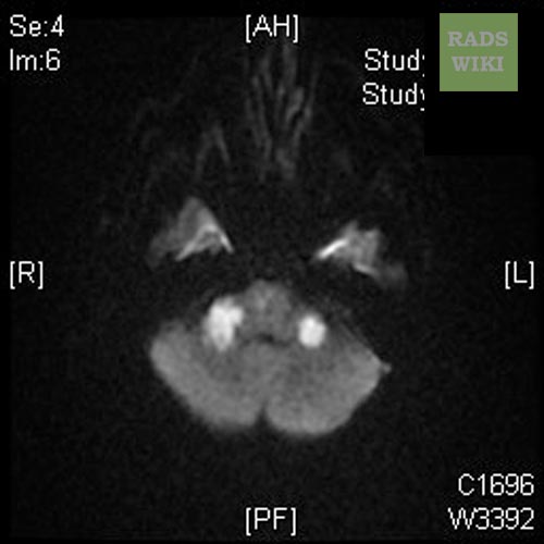
MRI - DWI
-

MRI - FLAIR


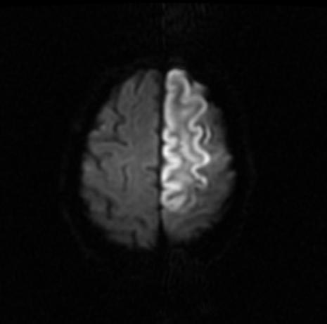
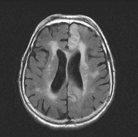
Patient No 2: Left ACA Infarction
-

MRI - T2
-

MRI - T1
-

MRI - ADC


-
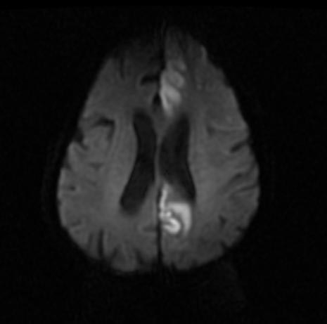
MRI- DWI
-

MRI- DWI


-

MRI- DWI
-
MRI- DWI


-
MRI- FLAIR
-

MRI- FLAIR


-
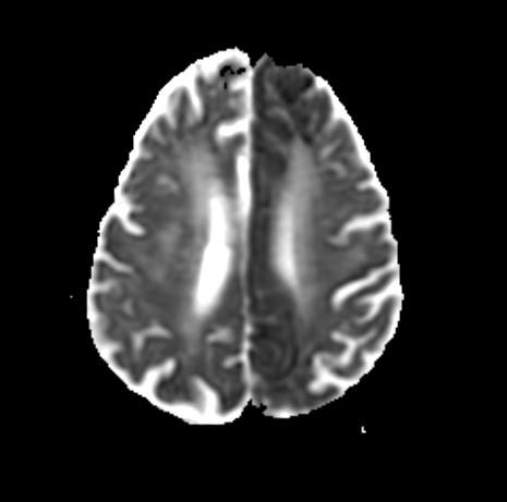
MRI- ADC
-
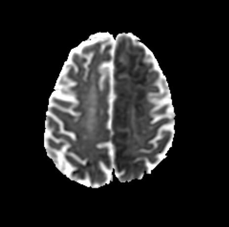
MRI- A


References
- ↑ Chalela, J (2007). "Magnetic resonance imaging and computed tomography in emergency assessment of patients with suspected acute stroke: a prospective comparison". Lancet. 369 (9558): 293–8. PMID 17258669. Retrieved 2008-01-22. Unknown parameter
|coauthors=ignored (help) - ↑ Kidwell, C (2004). "Comparison of MRI and CT for detection of acute intracerebral hemorrhage". JAMA. 292 (15): 1823–30. PMID 15494579. Retrieved 2008-01-22. Unknown parameter
|coauthors=ignored (help)