Intravenous leiomyomatosis
Editor-In-Chief: C. Michael Gibson, M.S., M.D. [1] Associate Editor-In-Chief: Cafer Zorkun, M.D., Ph.D. [2]; Ammu Susheela, M.D. [3]
Synonyms and keywords: Nesidioblastoma, IVLM
Overview
Intravenous leiomyomatosis is characterized by the extension into venous channels of histologically benign smooth muscle tumor arising from either the wall of a vessel or from a uterine leiomyoma. The etiology of intravenous leiomyomatosis is unclear. Intravenous leiomyomatosis must be differentiated from other diseases such as renal malignancies and sarcoma. The median age is 45 years, with patients ranging from 26 to 70 years old. Only females may develop intravenous leiomyomatosis. Common complications of intravenous leiomyomatosis include embolization, recurrence of the tumor, and metastasis. Surgery is the mainstay of therapy for intravenous leiomyomatosis.
Historical Perspective
- [Disease name] was first discovered by [scientist name], a [nationality + occupation], in [year] during/following [event].
- In [year], [gene] mutations were first identified in the pathogenesis of [disease name].
- In [year], the first [discovery] was developed by [scientist] to treat/diagnose [disease name].
Pathophysiology
- Intravenous leiomyomatosis is characterized by the extension into venous channels of histologically benign smooth muscle tumor arising from either the wall of a vessel or from a uterine leiomyoma.
- Patients are exclusively female, and the majority are white, premenopausal, and parous.
- Intravenous leiomyomatosis should be considered in young women with cardiac symptoms who have a right atrial mass as well as a pelvic mass or who have previously undergone hysterectomy for leiomyoma uterus with intravenous involvement.
Causes
- The etiology of intravenous leiomyomatosis is unclear.
Differentiating Intravenous Leiomyomatosis from other Diseases
- Intravenous leiomyomatosis must be differentiated from other diseases such as:
- Renal malignancies
- Sarcoma
- Thrombosis of the intravenous catheter
Epidemiology and Demographics
- The median age is 45 years, with patients ranging from 26 to 70 years old.
- Female are exclusively affected with intravenous leiomyomatosis.
- The prevalence of [disease name] is approximately [number or range] per 100,000 individuals worldwide.
- In [year], the incidence of [disease name] was estimated to be [number or range] cases per 100,000 individuals in [location].
Age
- Patients of all age groups may develop [disease name].
- [Disease name] is more commonly observed among patients aged [age range] years old.
- [Disease name] is more commonly observed among [elderly patients/young patients/children].
Gender
- [Disease name] affects men and women equally.
- [Gender 1] are more commonly affected with [disease name] than [gender 2].
- The [gender 1] to [Gender 2] ratio is approximately [number > 1] to 1.
Race
- There is no racial predilection for [disease name].
- [Disease name] usually affects individuals of the [race 1] race.
- [Race 2] individuals are less likely to develop [disease name].
Risk Factors
- Common risk factors in the development of [disease name] are [risk factor 1], [risk factor 2], [risk factor 3], and [risk factor 4].
Natural History, Complications and Prognosis
- The majority of patients with [disease name] remain asymptomatic for [duration/years].
- Early clinical features include [manifestation 1], [manifestation 2], and [manifestation 3].
- If left untreated, [#%] of patients with [disease name] may progress to develop [manifestation 1], [manifestation 2], and [manifestation 3].
- Common complications of [disease name] include [complication 1], [complication 2], and [complication 3].
- Prognosis is generally [excellent/good/poor], and the [1/5/10year mortality/survival rate] of patients with [disease name] is approximately [#%].
- Common complications of intravenous leiomyomatosis include embolization, recurrence of the tumor, and metastasis.
- The tumor is slow growing, and the prognosis is favorable.
Diagnosis
Diagnostic Criteria
- The diagnosis of [disease name] is made when at least [number] of the following [number] diagnostic criteria are met:
- [criterion 1]
- [criterion 2]
- [criterion 3]
- [criterion 4]
Symptoms
Patients may be asymptomatic or have symptoms of uterine leiomyomas, syncopal episodes, and dyspnea on exertion.
- [Disease name] is usually asymptomatic.
- Symptoms of [disease name] may include the following:
- [symptom 1]
- [symptom 2]
- [symptom 3]
- [symptom 4]
- [symptom 5]
- [symptom 6]
Physical Examination
- Patients with [disease name] usually appear [general appearance].
- Physical examination may be remarkable for:
- [finding 1]
- [finding 2]
- [finding 3]
- [finding 4]
- [finding 5]
- [finding 6]
Laboratory Findings
- There are no specific laboratory findings associated with [disease name].
- A [positive/negative] [test name] is diagnostic of [disease name].
- An [elevated/reduced] concentration of [serum/blood/urinary/CSF/other] [lab test] is diagnostic of [disease name].
- Other laboratory findings consistent with the diagnosis of [disease name] include [abnormal test 1], [abnormal test 2], and [abnormal test 3].
Imaging Findings
- There are no [imaging study] findings associated with [disease name].
- [Imaging study 1] is the imaging modality of choice for [disease name].
- On [imaging study 1], [disease name] is characterized by [finding 1], [finding 2], and [finding 3].
- [Imaging study 2] may demonstrate [finding 1], [finding 2], and [finding 3].
Other Diagnostic Studies
- [Disease name] may also be diagnosed using [diagnostic study name].
- Findings on [diagnostic study name] include [finding 1], [finding 2], and [finding 3].
Treatment
Medical Therapy
- There is no treatment for [disease name]; the mainstay of therapy is supportive care.
- The mainstay of therapy for [disease name] is [medical therapy 1] and [medical therapy 2].
- [Medical therapy 1] acts by [mechanism of action 1].
- Response to [medical therapy 1] can be monitored with [test/physical finding/imaging] every [frequency/duration].
Surgery
- Surgery is the mainstay of therapy for [disease name].
- [Surgical procedure] in conjunction with [chemotherapy/radiation] is the most common approach to the treatment of [disease name].
- [Surgical procedure] can only be performed for patients with [disease stage] [disease name].
Prevention
- There are no primary preventive measures available for [disease name].
- Effective measures for the primary prevention of [disease name] include [measure1], [measure2], and [measure3].
- Once diagnosed and successfully treated, patients with [disease name] are followed-up every [duration]. Follow-up testing includes [test 1], [test 2], and [test 3].
References
Images
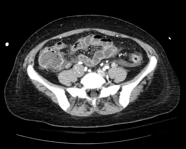
Example #1
The patient presented with S.O.B. one year after hysterectomy for a leiomyomatous uterus.
-
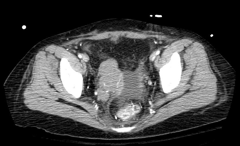
CT in Intravenous leiomyomatosis
-


Treatment
- Surgery is the mainstay of therapy for intravenous leiomyomatosis.
Related Chapters
- Uterine leiomyoma
- Benign metastasizing leiomyoma