Hydronephrosis: Difference between revisions
No edit summary |
|||
| Line 56: | Line 56: | ||
<gallery> | <gallery> | ||
Image:Hydronephrosis 001.jpg | Image:Hydronephrosis 001.jpg | ||
Image:Hydronephrosis 003.jpg | Image:Hydronephrosis 003.jpg | ||
</gallery> | </gallery> | ||
Revision as of 22:32, 14 March 2009
| Hydronephrosis | |
 | |
|---|---|
| Ultrasound picture of hydronephrosis caused by a left ureteral stone | |
| ICD-10 | N13.0-N13.3 |
| ICD-9 | 591 |
| DiseasesDB | 6145 |
| MeSH | D006869 |
Template:Search infobox Steven C. Campbell, M.D., Ph.D.
Contributors: Cafer Zorkun M.D., PhD.
Please Join in Editing This Page and Apply to be an Editor-In-Chief for this topic: There can be one or more than one Editor-In-Chief. You may also apply to be an Associate Editor-In-Chief of one of the subtopics below. Please mail us [1] to indicate your interest in serving either as an Editor-In-Chief of the entire topic or as an Associate Editor-In-Chief for a subtopic. Please be sure to attach your CV and or biographical sketch.
Overview
Hydronephrosis is distention and dilation of the renal pelvis, usually caused by obstruction of the free flow of urine from the kidney.[1]
Etiology
The obstruction may be either partial or complete and can occur anywhere from the urethral meatus to the calyces of the renal pelvis.
The obstruction may arise from either inside or outside the urinary tract or may come from the wall of the urinary tract itself. Intrinsic obstructions (those that occur within the tract) include blood clots, stones, sloughed papilla along with tumours of the kidney, ureter and bladder. Extrinsic obstructions (those that are caused by factors outside of the urinary tract) include pelvic or abdominal tumours or masses, retroperitoneal fibrosis or neurological deficits. Strictures of the ureters (congenital or acquired), neuromuscular dysfunctions or schistosomiasis are other causes which originate from the wall of the urinary tract.
Signs and symptoms
The signs and symptoms of hydronephrosis depends upon whether the obstruction is acute or chronic. Unilateral hydronephrosis may even occur without symptoms.[1]
Blood tests can show raised creatinine and electrolyte imbalance. Urinalysis may show an elevated pH due to the secondary destruction of nephrons within the affected kidney.
Symptoms that occur regardless of where the obstruction lies include loin or flank pain. An enlarged kidney may be palpable on examination.
Where the obstruction occurs in the lower urinary tract, suprapubic tenderness (with or without a history of bladder outflow obstruction) along with a palpable bladder are strongly suggestive of acute urinary retention, which left untreated is highly likely to cause hydronephrosis.
Upper urinary tract obstruction is characterised by pain in the flank, often radiating to either the abdomen or the groin. Where the obstruction is chronic, renal failure may also be present. If the obstruction is complete, an enlarged kidney is often palpable on examination.
Diagnosis
Prenatal diagnosis is possible.[2]
Blood (U&E, creatinine) and urine (MSU, pH) tests should be taken. IVUs, ultrasounds, CTs and MRIs are also important tests. Ultrasound allows for visualisation of the ureters and kidneys and can be used to assess the presence of hydronephrosis and/or hydroureter. An IVU is useful for assessing the position of the obstruction. Antegrade or retrograde pyelography will show similar findings to an IVU but offer a therapeutic option as well.
The choice of imaging depends on the clinical presentation (history, symptoms and examination findings). In the case of renal colic (one sided loin pain usually accompanied by a trace of blood in the urine) the initial investigation is usually an intravenous urogram. This has the advantage of showing whether there is any obstruction of flow of urine causing hydronephrosis as well as demonstrating the function of the other kidney. Many stones are not visible on plain xray or IVU but 99% of stones are visible on CT and therefore CT is becoming a common choice of initial investigation. MRI is less commonly used, often when there is a reason to avoid radiation exposure, e.g. in pregnancy.
Patient #1: CT images demonstrate marked left hydronephrosis secondary to a calculus in the proximal right ureter


Ultrasonography
-
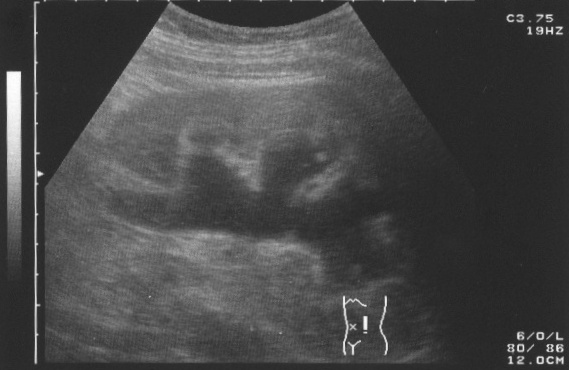
Ultrasound picture of hydronephrosis caused by a left ureteral stone
Histopathological Fidings
-
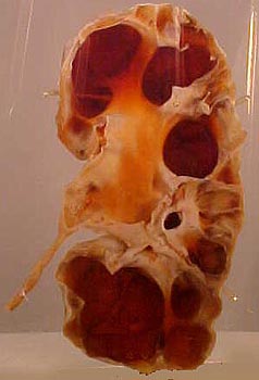
Specimen of a kidney that has undergone extensive dilation due to hydronephrosis. Note the extensive atrophy and thinning of the renal cortex.

Treatment
Treatment of hydronephrosis focuses upon the removal of the obstruction and drainage of the urine that has accumulated behind the obstruction. Therefore, the specific treatment depends upon where the obstruction lies, and whether it is acute or chronic.
Acute obstruction of the upper urinary tract is usually treated by the insertion of a nephrostomy tube. Chronic upper urinary tract obstruction is treated by the insertion of a ureteric stent or a pyeloplasty.
Lower urinary tract obstruction (such as that caused by bladder outflow obstruction secondary to prostatic hypertrophy) is usually treated by insertion of a urinary catheter or a suprapubic catheter.
Surgery is not required in all cases.[3]
Complications
Left untreated, bilateral obstruction (obstruction occurring to both kidneys rather than one) has a poor prognosis.
References
- ↑ 1.0 1.1 Kumar, Vinay; Fausto, Nelson; Fausto, Nelso; Robbins, Stanley L.; Abbas, Abul K.; Cotran, Ramzi S. (2005). Robbins and Cotran Pathologic Basis of Disease (7th ed.). Philadelphia, Pa.: Elsevier Saunders. pp. 1012–1014. ISBN 0-7216-0187-1.
- ↑ Estrada CR (2008). "Prenatal hydronephrosis: early evaluation". Curr Opin Urol. 18 (4): 401–3. doi:10.1097/MOU.0b013e328302edfe. PMID 18520762. Unknown parameter
|month=ignored (help) - ↑ Onen A (2007). "Treatment and outcome of prenatally detected newborn hydronephrosis". J Pediatr Urol. 3 (6): 469–76. doi:10.1016/j.jpurol.2007.05.002. PMID 18947797. Unknown parameter
|month=ignored (help)
External Links
See also
Template:Nephrology Template:SIB