Hepatocellular adenoma MRI
|
Hepatocellular adenoma Microchapters |
|
Diagnosis |
|---|
|
Treatment |
|
Case Studies |
|
Hepatocellular adenoma MRI On the Web |
|
American Roentgen Ray Society Images of Hepatocellular adenoma MRI |
|
Risk calculators and risk factors for Hepatocellular adenoma MRI |
Editor-In-Chief: C. Michael Gibson, M.S., M.D. [1]; Associate Editor(s)-In-Chief: Cafer Zorkun, M.D., Ph.D. [2]Nawal Muazam M.D.[3]
Overview
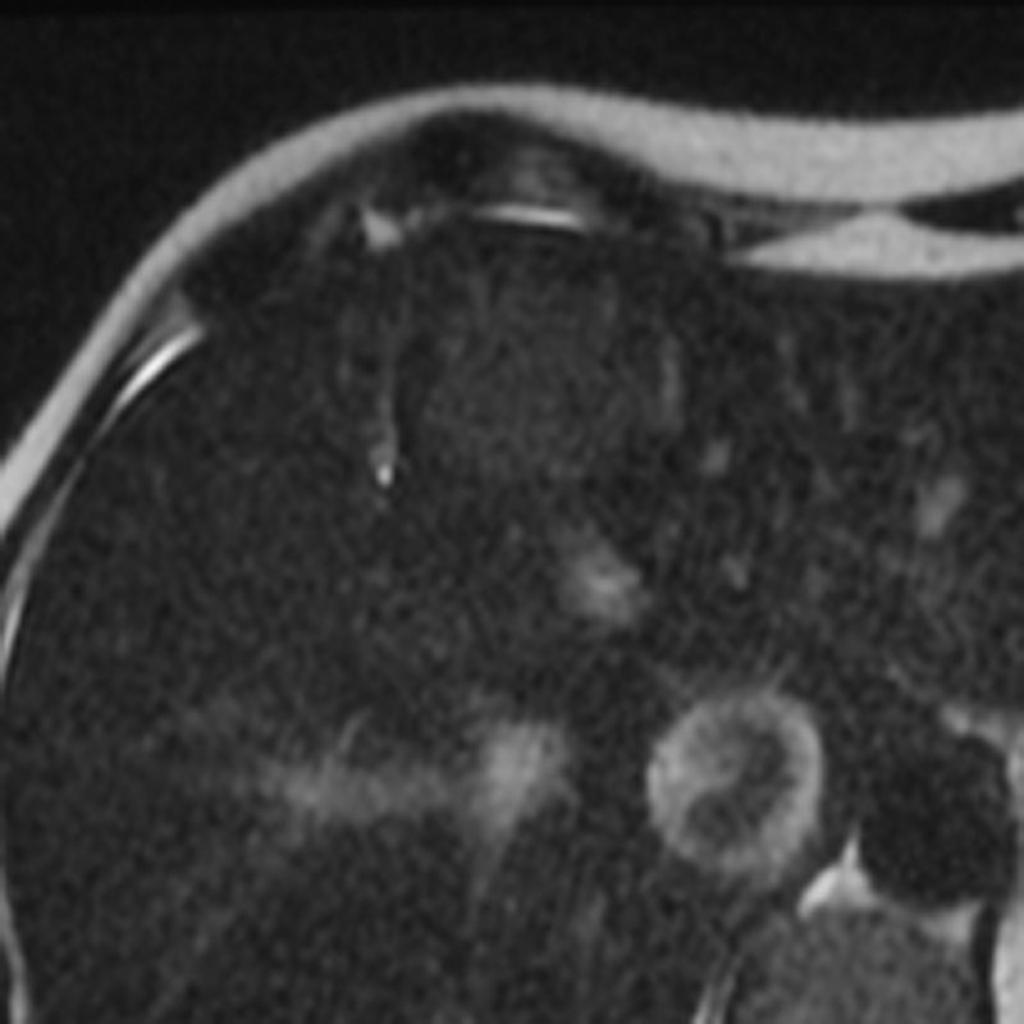
The MRI findings of hepatocellular adenoma include; from hyperintense to mildly hypointense relatvive to the liver tissue on T1 weighted images. On T2 weighted images they are predominantly hyperintense relative to the liver, whereas in the presence of necrosis and hemorrhage they can be heterogeneous with hyper or hypo attenuating signal. The central scar is on godalinium is not seen in hepatocellular adenoma. There is usually no significant uptake with injection of hepatocellular specific contrast agent, godalinium benzoyloxypropionictetraacetate (Gd-BOTA).
MRI
- The MR imgaing findings of hepatocellualar adenoma include;
- From hyperintense to mildly hypointense relative to liver tissue on T1 weighted images. This heterogeneous appearance results from areas of high intensity due to fat and acute hemorrhage and low signal intensity area corresponding to necrosis or old hemorrhage or calcifications.
- The hepatocellular adenoma in T2 weighted images are predominantly hyperintense relative to liver, although in the presence of necrosis and hemorrhage they can be heterogeneous with hyper and hypo attenuating signal.
- Dynamic postgodalinium show intense arterial phase enhancement with isointensity on portal phase and delayed images.
- Hepatocellular adenoma do not have a central scar so if a central scar enhances after godalinium is administered, the diagnosis of focal nodular hyperplasia is strongly favored.
- A central scar has never been reported in hepatocellular adenomas.
- With the injection of hepatocellular specific contrast agent, godalinium benzyloxypropionictetraacetate (Gd-BOTA) there is usually no significant uptake.
-
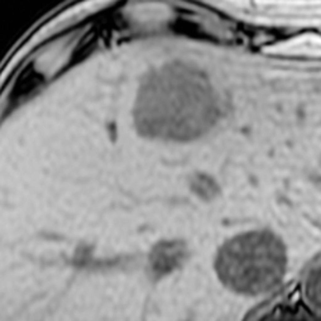
liver MRI show a large, subtle T2 hyperintense, well defined rounded lesion in the liver which demonstrates homogenous signal loss on T1 opposed phase scans.
-