Gestational trophoblastic neoplasia CT
|
Gestational trophoblastic neoplasia Microchapters |
|
Differentiating Gestational trophoblastic neoplasia from other Diseases |
|---|
|
Diagnosis |
|
Treatment |
|
Case Studies |
|
Gestational trophoblastic neoplasia CT On the Web |
|
American Roentgen Ray Society Images of Gestational trophoblastic neoplasia CT |
|
Directions to Hospitals Treating Gestational trophoblastic neoplasia |
|
Risk calculators and risk factors for Gestational trophoblastic neoplasia CT |
Editor-In-Chief: C. Michael Gibson, M.S., M.D. [1]Associate Editor(s)-in-Chief: Monalisa Dmello, M.B,B.S., M.D. [2]
Overview
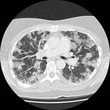
CT scan may be performed to detect metastasis of choriocarcinoma to lung, brain, and liver.
CT
CT scan may be performed to detect metastasis of choriocarcinoma to lung, brain, and liver. CT scan is used to check for the spread of choriocarcinoma outside the uterus.
-
A
-
B
-
C
-
D
-
E


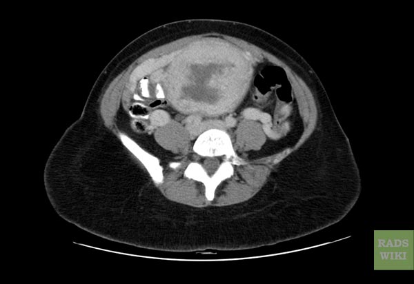
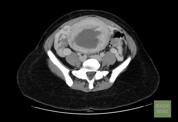
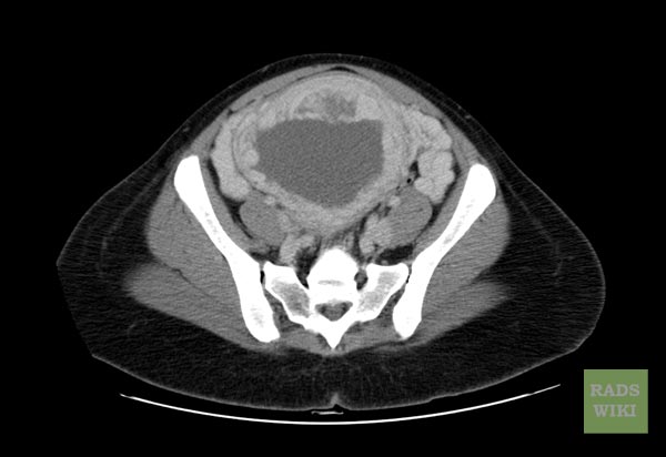
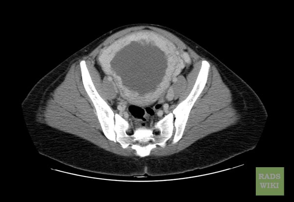
- Figures A-D: CT scan findings are consistent with choriocarcinoma (A-D, axial).[1]
- Figures E: CT showing patient of choriocarcinoma with lung metastasis.[2]
References
- ↑ Image courtesy of Radswiki. Radiopaedia (original file [ http://radiopaedia.org/articles/choriocarcinoma ‘’here’’]). Creative Commons BY-SA-NC
- ↑ Image courtesy of Dr Maxime St-Amant. Radiopaedia (original file [ http://radiopaedia.org/articles/choriocarcinoma ‘’here’’]). Creative Commons BY-SA-NC