Galeazzi fracture
.jpg)
|
WikiDoc Resources for Galeazzi fracture |
|
Articles |
|---|
|
Most recent articles on Galeazzi fracture Most cited articles on Galeazzi fracture |
|
Media |
|
Powerpoint slides on Galeazzi fracture |
|
Evidence Based Medicine |
|
Cochrane Collaboration on Galeazzi fracture |
|
Clinical Trials |
|
Ongoing Trials on Galeazzi fracture at Clinical Trials.gov Trial results on Galeazzi fracture Clinical Trials on Galeazzi fracture at Google
|
|
Guidelines / Policies / Govt |
|
US National Guidelines Clearinghouse on Galeazzi fracture NICE Guidance on Galeazzi fracture
|
|
Books |
|
News |
|
Commentary |
|
Definitions |
|
Patient Resources / Community |
|
Patient resources on Galeazzi fracture Discussion groups on Galeazzi fracture Patient Handouts on Galeazzi fracture Directions to Hospitals Treating Galeazzi fracture Risk calculators and risk factors for Galeazzi fracture
|
|
Healthcare Provider Resources |
|
Causes & Risk Factors for Galeazzi fracture |
|
Continuing Medical Education (CME) |
|
International |
|
|
|
Business |
|
Experimental / Informatics |
Editor-In-Chief: C. Michael Gibson, M.S., M.D. [1]; Associate Editor(s)-in-Chief: Mohammadmain Rezazadehsaatlou[2].
Overview[1][2]
The Galeazzi fracture-dislocation is an orthopedic injury pattern with the following definition:
- An isolated fractures of the distal 1/3 radius shaft
- Associated distal radioulnar joint (DRUJ) injury
Historical Perspective[3][4][5][6]
In 1822, Sir Astley Cooper worked on the dislocations and Fracture of human body.
In 1934, Riccardo Galeazzi , an Italian surgeon at the Instituto de Rachitici in Milan, reported on his experience with 18 fractures with with close similarities to the Monteggia lesion.
In 1941, Campbell termed the Galeazzi fracture the "fracture of necessity".
In 1957, Hughston presented the definitive management of the Galeazzi fracture.
Causes[6][7][8]
The main etiology of the Galeazzi fracture-dislocation is thought to be an axial loading may be placed on a hyperpronated forearm during falling onto an outstretched hand (FOOSH) with an extended wrist and hyperpronated forearm. Because at this posture the energy from the radius fracture transmitted towards the radioulnar joint cause the dislocation of the DRUJ.
Pathophysiology [6][1][2][7][8]
Mechanism
The Galeazzi fracture-dislocation is caused by a fall on the outstretched hands with the wrist in dorsiflexion position. The form and severity of this fracture depends on the position of the wrist at the moment of hitting the ground. The width of this mentioned angle affects the localization of the fracture. Pronation, supination and abduction positions leads the direction of the force and the compression of carpus and different appearances of injury.
Pathophysiology [9][10]
Its known that the Galeazzi fracture-dislocation in normal healthy adults can be caused due to the high-energy trauma (e.g., motor vehicle accidents), sport related injuries, falling from height. But it should be noted that the most important Risk factors for insufficiency fractures is chronic metabolic disease such as steoporosis, osteopenia, eating-disordered behavior, higher age, prolonged corticosteroid usage, female gender, lower BMI, history of a recent falling, and prior fracture.
- The pattern of bone fracture and severity of injury depends on variety of factors such as:
- Patients age
- Patients Weight
- Patients past medical history specifically any bone diseases affecting the quality of bone (such as osteoporosis, malignancies)
- Energy of trauma
- Bone quality
- Position of the specific organ during the trauma
- The below-mentioned processes cause decreased bone mass density:
- Autophagy is the mechanism through which osteocytes evade oxidative stress.
- The capability of autophagy in cells decreases as they age, a major factor of aging.
- As osteocytes grow, viability of cells decrease thereby decreasing the bone mass density.
Differentiating Galeazzi fracture-dislocation from other Diseases[11][12][9][10]
In the orthopedic medicine its important to know that the forearm fracture should be evaluated using radiography for both confirming diagnosis and also for evaluating the surrounding tissues. Other injuries such as possible olecranon fracture-dislocation; radial head or coronoid fractures or lateral collateral ligament injury, might be seen in Monteggia fracture. If the mechanism of injury suggests particularly low energy then the Osteoporosis should be considered. The pathological Fractures occurring in a bone with a tumor or Paget's disease) are rare but possible[3].
Also it should be noted that the both bone fractures can be complicated by acute compartment syndrome of the forearm. Signs suggesting compartment syndrome are pain on extension of digits, and marked edema[3].
As another important fact in orthopedic fracture is if both-bone fractures were found in pediatric which is common after accidental trauma, but it may also be the due to the of child abuse; and in these cases a careful attention and evaluation should be considered if a child abuse is suspected
Epidemiology and Demographics [13]
Galeazzi fractures account for around 3-7% of all forearm fractures in adults. Normally, 25% of the radial shaft fractures are true Galeazzi type injuries. The most common risk factors for the Galeazzi fracture are: sports (football and wrestling), osteoporosis, and post-menopausal time; consequently, These risk factors cause the highest occurrence in young males (10:10,000) and elderly females (5:10,000). The peak incidence in children is the age of 9 to 12.
Risk Factors[14][15][16]
There are different risk factors that presidpose patient for the Galeazzi fracture-dislocation that include:
- High-risk contact sports
- Higher age (elderly adults are higher prone to such fractures)
- Reduced bone density (osteoporosis)
- Direct blow
- Road / traffic accidents
- Falling on an outstretched hand with the forearm pronated.
- Direct trauma to the arm/forearm
- Taking part in any rough or high-impact sport
- Street fights, gunshot wounds, and domestic violence, may also cause the Galeazzi fracture-dislocation
- Road traffic accidents.
Screening[17][18][19][20]
Osteoporosis is an important risk factor for human affecting human bone especially in men with the age of older than 50 years old and postmenopausal and women.
Based on the US Preventive Services Task Force (USPSTF) there are three groups of patients need to be screened for the osteoporosis:
- · Men with no history of osteoporosis
- · Women with the age of 65≤ year old, with no previous history of pathological fracture due to the osteoporosis
- · Women with the age of <65 years, with 10-year fracture risk of not less than a 65-year-old white woman (who has not any other risk factor)
Accordingly women older than age of 50 are the main target for the osteoporosis screening. There is no specific recommendation to screen men for the osteoporosis.
The USPSTF recommendations from 2002 included:
Meanwhile, there are two major modalities for the osteoporosis screening:
- · Dual energy x-ray absorptiometry (DXA) of the hip and lumbar spine bones
- · Quantitative ultrasonography of the calcaneus
*It should be noted of the two above mentioned modalities for screening the ultrasonograhy is preferred to the DXA due to its lower cost, lower ionizing radiation, more availability.
After the primary evaluation of the osteoporosis, the further evaluation are required in some cases such as:
· Women with normal bone density or mild osteopenia: T-score of greater than −1.50 – should have screening for 15 years.
· Women with moderate osteopenia: T-score of −1.50 to −1.99 – should have screening for 5 years.
· Women with advanced osteopenia: T-score of −2.00 to −2.49 - should have screening for 1 year.
Natural History, Complications and Prognosis [21][22]
Natural History
In cases with untreated Galeazzi fracture-dislocation the malunion and deformity of arm can be occurred.
Complications
The overall complication rate in the treatment of Galeazzi fracture-dislocation were found in around 40% of cases:
- Neurovascular compromise: such as Ulna nerve damage
- Compartment syndrome
- Chronic disability of the DRUJ
- Physeal Injury
- Malunion of the radius
- Nonunion
- Infection
- Refracture following plate removal
- Posterior interosseois nerve (PIN) injury.
- Instability of the DRUJ
Prognosis
Successful treatment of Galeazzi fracture-dislocation depends on the on-time interventions such as: reduction of the radius and DRUJ and the restoration of the forearm axis. The incidence of nonunion of Galeazzi fracture-dislocation is very low. On the other hand, the rate of successful union following the open reduction of forearm fractures was reported around 98%. Previous researches showed that the loss of strength at the supination and pronation were found in 12.5% and 27.2%, respectively.
Diagnosis[23][24][25][26]
The diagnosis of a Galeazzi fracture-dislocation should be confirmed using a radiographic examination.
Accordingly, related classification is based on the position of the radius:
Type I
- Dorsal displacement of the radius (Common type)
- Caused by supination force
Type II
- Volar displacement of the radius (Rare type)
- Caused by pronation force
The two main views such as anteroposterior (AP) and lateral forearm are needed in this regard:
- Radial shaft fracture:
- Commonly found at the junction of the middle and distal third
- Dorsal/Volar angulation
- Radial shortening may occur
- Dislocation of the distal radioulnar joint
Meanwhile, the following mentioned findings on the obtained radiography (such as plain radiography and the bilateral axial computed tomography (CT)) are suggestive of injury to the distal radioulnar joint (DRUJ):
- The dislocated radius near to the injury site
- Shortened radius by more than 5 mm near to the injury site
- The ulnar styloid base fracture near to the injury site
- Widening of the DRUJ space near to the injury site
History and Symptoms[26][27]
The related signs and symptoms include:
- Deformity
- Skin lacerations
- Weak pulse
- Open fractures
- Bruising
- Swelling
- Stiffness
- Inability to move
- Pain in touch
- Loss of function of the forearm
- Difficulties in detection of pulses
- Radial nerve damage
In the physical exam the orthopedic surgeon should check the vascular status and amount of swelling in the forearm. In MULTI-trauma patients or in comatose or obtunded patients a tense compartment with neurological signs or stretch pain should be considered as the compartment syndrome, and the compartment pressures should be measured and monitored. Normally the pain and soft-tissue swelling are found at the injury site (distal-third radial fracture site and at the wrist joint). This injury should be confirmed using a radiographic evaluations. Also, patients may loss the pinch mechanism between their thumb and their index finger which can be due to the paralysis of the flexor pollicis longus (FPL) and flexor digitorum profundus (FDP).
Physical Examination[28]
The related signs and symptoms include:
- Edema of the forearm
- Most of the time the edema will be a non-pitting edema
- Depends on the edema extent, it may even lead to compartment syndrome in the anterior and internal compartment of forearm
- Bruising
- As a manifestation of internal injury to the local vessels by trauma or fractures bone
- Decrease in range of motion
- Movement of the fractures limb will be painful if possible at all
- Tenderness
- Deformity
- Fractured bone deformity may be touchable in the internal side of the forearm if the fracture is displaced
In the physical exam the orthopedic surgeon should check the vascular status and amount of swelling in the forearm. In polytrauma patients or in comatose or obtunded patients a tense compartment with neurological signs or stretch pain should be considered as the compartment syndrome, and the compartment pressures should be measured and monitored.
Physical examination of patients with Galeazzi fracture-dislocation is usually remarkable for swelling, tenderness, bruises, ecchymosis, deformity and restricted range of motion of the wrist.
Appearance of the Patient [29][30]
- Patients with Galeazzi fracture-dislocation usually appears normal unless the patients had a high energy trauma causing the open wound fracture.
Vital Signs
- Weak pulse may be seen when associated with polytrauma.
- Low blood pressure with normal pulse pressure may be present due to compound fracture with blood loss.
Skin
- Skin examination of patients withGaleazzi fracture-dislocation includes:
HEENT
- HEENT examination of patients with Galeazzi fracture-dislocation is usually normal.
Neck
- Neck examination of patients with Galeazzi fracture-dislocation is usually normal
Lungs
- Pulmonary examination of patients with Galeazzi fracture-dislocation is usually normal
Heart
- Cardiovascular examination of patients with Galeazzi fracture-dislocation is usually normal
Abdomen
- Abdominal examination of patients with Galeazzi fracture-dislocation is usually normal
Back
- Back examination of patients with Galeazzi fracture-dislocation is usually normal
Genitourinary
- Genitourinary examination of patients with Galeazzi fracture-dislocation is usually normal
Neuromuscular
- Neuromuscular examination of patients with Galeazzi fracture-dislocation is usually normal
- However, some patients may develop neuropraxia of the branch of the Ulnar nerve resulting in decreased sensation of thumb, index and middle finger.
Laboratory Findings[30]
There is a limited laboratory tests useful in the diagnosis of bone fractures such as the Galeazzi fracture-dislocation. Meanwhile, aged men and women may have some abnormalities in their laboratory findings suggestive of osteoporosis.
Laboratory tests for the diagnosis of osteoporosis are:
- Complete blood count (CBC)
- Serum total calcium level
- Serum Ionized calcium level
- Serum phosphate level
- Serum alkaline phosphatase level
- Serum 25-(OH)-vitamin D level
X Ray[23][24][25]
The orthopedic surgeon should consider to have at least two radiographic projections (ie, anteroposterior [AP] and lateral) of the forearm. These show the fracture, the extent of displacement, and the extent of comminution. The orthopedic surgeon should pay serious attention toward finding any foreign bodies in open fractures and gunshot injuries. Also imperative is to include the elbow and wrist joint in the radiographs of Galeazzi fracture-dislocation to ensure that the distal radioulnar joint injuries are not missed.
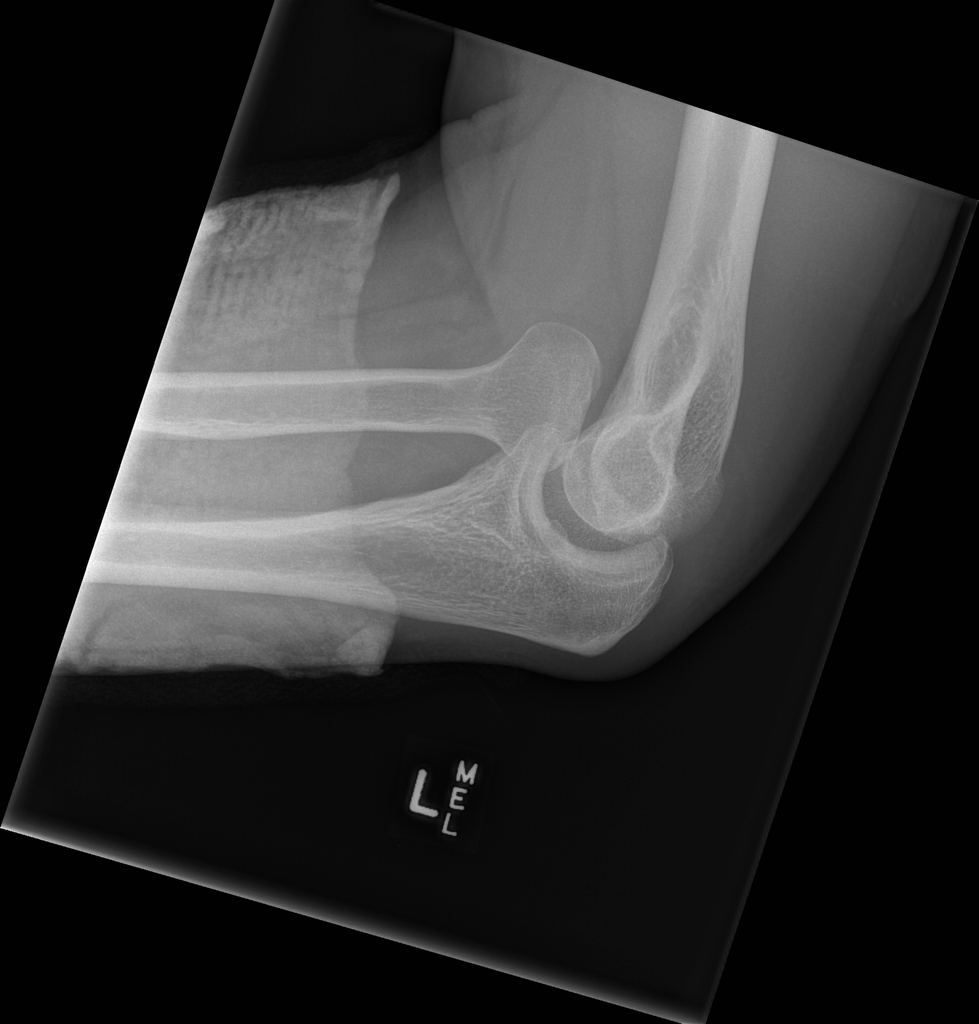
-
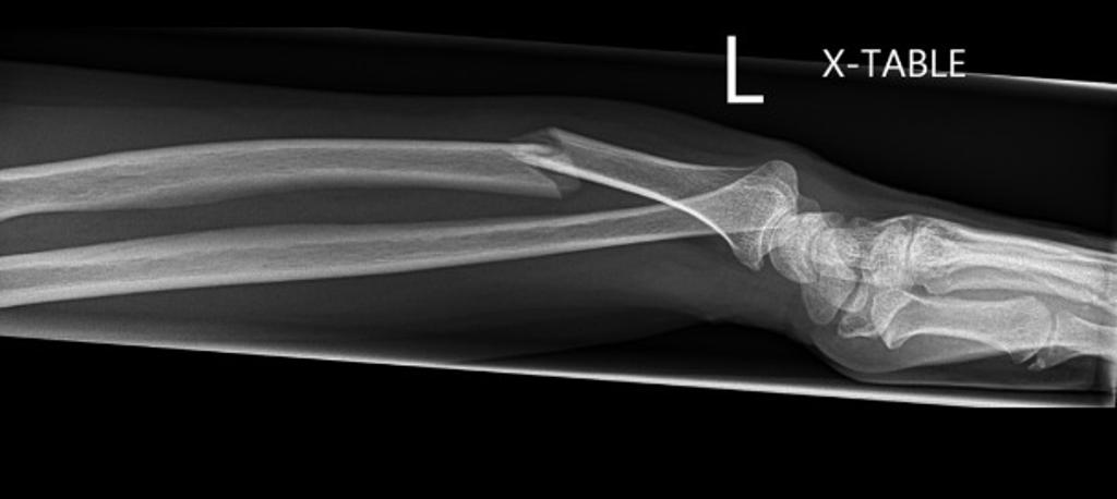
Lateral- Type 1 Galeazzi fracture
-
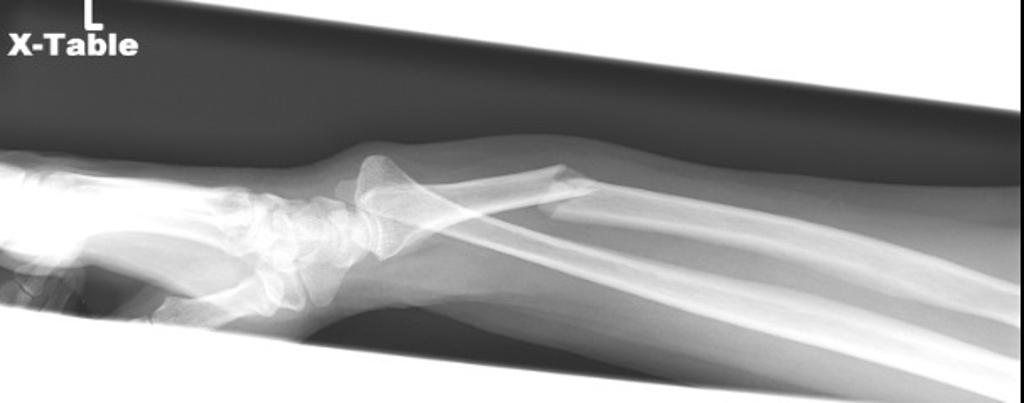
Lateral- Type 1 Galeazzi fracture
-
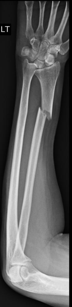
PA- Type 1 Galeazzi fracture
-
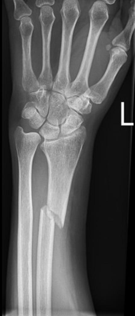
PA- Type 1 Galeazzi fracture
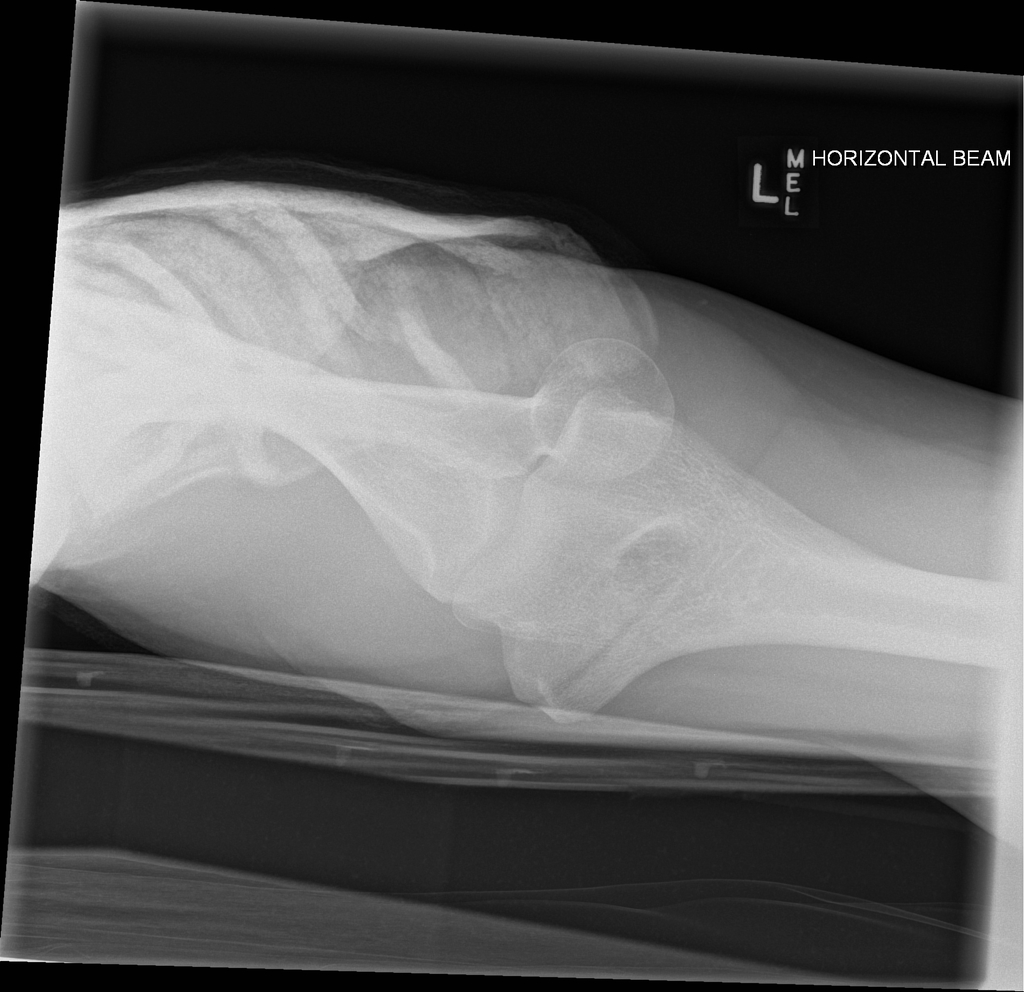
-
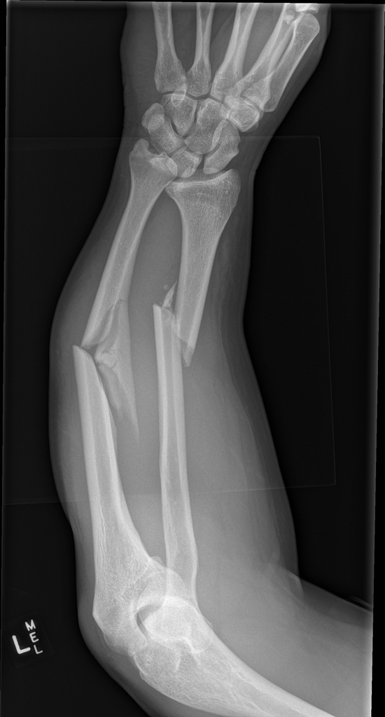
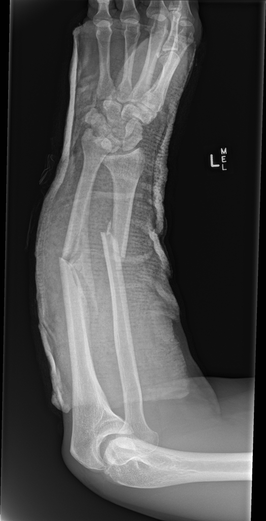
Combined Monteggia and Galeazzi fractures- Comminuted fractures of the ulnar and radial midshafts with significant radial and volar angulation. Large ulnar butterfly fragment. Dorsally dislocated ulnar head (DRUJ). Abnormal radiocapellar alignment which requires formal elbow views.
-
Combined Monteggia and Galeazzi fractures- Comminuted fractures of the ulnar and radial midshafts with significant radial and volar angulation. Large ulnar butterfly fragment. Dorsally dislocated ulnar head (DRUJ). Abnormal radiocapellar alignment which requires formal elbow views.

.jpg)


CT [31]
- CT-scan in the case of the Galeazzi fracture-dislocation is the best modality if you can not have an exclusive diagnosis by X-ray itself can not be made.
MRI [30]
- Magnetic resonance imaging (MRI) is an expensive technique that should not be used routinely.
- MRI is a powerful diagnostic tool to assess the abnormalities of the bone, ligaments and soft tissues associated with the Galeazzi fracture-dislocation, but it is known as a limited utility in radioulnar injuries and is not indicated in uncomplicated forearm fractures.
- Meanwhile, the MRI can be useful in in following mentioned evaluations:
- Evaluation of occult fractures
- Evaluation of the post-traumatic or avascular necrosis of carpal bones
- Evaluation of tendons
- Evaluation of nerve
- Evaluation of carpal tunnel syndrome
Other Imaging Findings[9][13]
There are no other imaging findings associated with Galeazzi fracture-dislocation
Other Diagnostic Studies[9][13]
There are no other Diagnostic studies associated with Galeazzi fracture-dislocation
Treatment[29]
Immediate stabilization of patients is the first step. Then the radial fracture and the DRUJ stabilization is recommended in these cases. Open forearm fractures considered as a surgical emergency. Galeazzi fractures occurs in younger patients who are skeletally immature; the normally they treated using a closed reduction and casting. Since closed reduction and cast application have led to unsatisfactory results. Then, Almost always the open reduction are necessary for the Galeazzi fractures. There are controversies regarding the indications for intramedullary nailing of forearm fractures.
Non-Operative Treatment[29]
- The first step in managing a patient with a fracture is to stabilize the patient if he/she is unstable due to blood loss, etc by giving them intravenous fluids and giving them some painkillers if the pain is severe.
- In children, the usual plan is to attempt closed reduction followed by cast immobilization. In adults, treatment with immobilization in a molded long arm cast can be used in those rare occasions of a non-displaced fracture of both bones of the forearm. If the fracture shifts in position, it may require surgery to put the bones back together.
- Rigid immobilization is suggested in preference to removable splints in nonoperative treatment for the management of the Galeazzi fracture-dislocation
- For all patients with Galeazzi fracture-dislocation, a post-reduction true lateral radiograph is suggested .
- Operative fixation is suggested in preference to cast fixation for fractures with post-reduction radial shortening greater than 3 mm, dorsal tilt greater than 10º, or intra-articular displacement or step-off greater than 2 mm.
- Patients probably do not need to begin early wrist motion routinely after stable fracture fixation.
- Adjuvant treatment of Galeazzi fracture-dislocation with vitamin C is suggested for the prevention of disproportionate pain
-
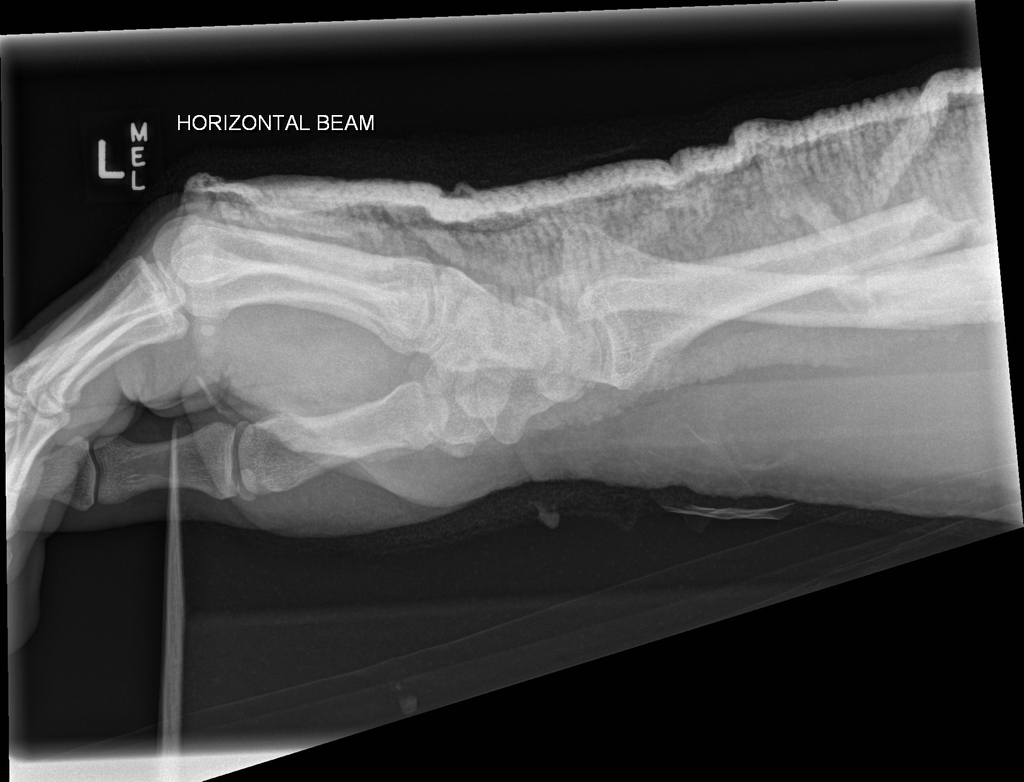
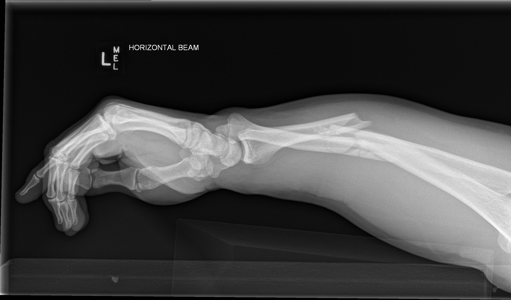
Combined Monteggia and Galeazzi fractures- Short arm backslab. Ulnar and radial midshaft fractures maintain similar alignment. Dorsally dislocated ulnar head (DRUJ). Volarly dislocated radial head (PRUJ) confirmed. Lat view
-
Combined Monteggia and Galeazzi fractures- Short arm backslab. Ulnar and radial midshaft fractures maintain similar alignment. Dorsally dislocated ulnar head (DRUJ). Volarly dislocated radial head (PRUJ) confirmed. AP view
-
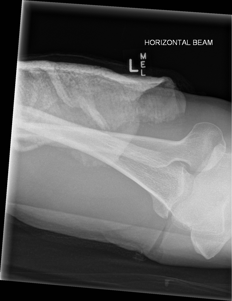
Combined Monteggia and Galeazzi fractures- Short arm backslab. Ulnar and radial midshaft fractures maintain similar alignment. Dorsally dislocated ulnar head (DRUJ). Volarly dislocated radial head (PRUJ) confirmed. AP view


Complications of Non-surgical therapy [9][29][13]
Failure of non-surgical therapy is common:
- Re-displacement to its original position even in a cast
- Stiffness
- Post traumatic osteoarthritis leading to wrist pain and loss of function
- Other risks specific to cast treatment include:
- Compression of the swollen arm causing compartment syndrome or carpal tunnel syndrome
- Reflex sympathetic dystrophy is a serious complication
- Stiffness is universal following a prolonged period of immobilization and swelling
Surgery[9] [13][9][13]
Returning to the normal physical activity after Galeazzi fracture-dislocation can take weeks to months of therapy under supervision an orthopedist. Meanwhile, a physiotherapy can be helpful for patient to achieve the normal wrist and elbow function caused by the immobilisation. All adult Galeazzi fractures should be considered to be treated with open reduction and internal fixation (ORIF).
-
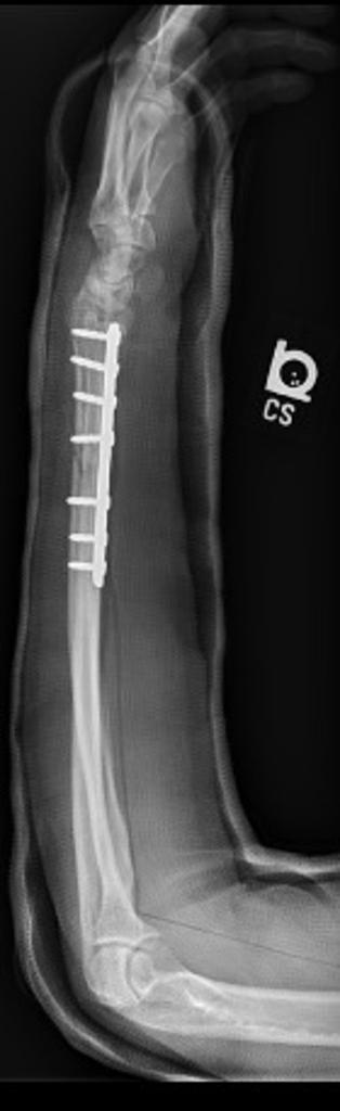
PA- Type 1 Galeazzi fracture
Operation [29]</nowiki>
- There are a variety of methods and implants useful to stabilize the Galeazzi fracture-dislocation, ranging from closed reduction and percutaneous pin fixation to the use of intra-medullary devices.
- However, the most common fixation methods to treat complex Galeazzi fracture-dislocation include external fixation, and open reduction and internal fixation.
External Fixation With or Without Percutaneous Pin Fixation
- Wrist spanning external fixation employs ligamentotaxis to restore and maintain length, alignment, and rotation of ulnar bone.
- Reduction is typically obtained through closed or minimally open methods and preserves the fracture biology.
- The addition of percutaneous pins enhances the ability to reduce and stabilize fracture fragments.
Complications of External Fixation [9][13]
- Pin tract infection
- Injury to the superficial branch of the nerve
- Complex regional pain syndrome
Open reduction and internal fixation with plates and screws [32]
- This is the most common type of surgical repair for Galeazzi fracture-dislocation
- During this type of procedure, the bone fragments are first repositioned (reduced) into their normal alignment.
- The bones held together with special screws and metal plates attached to the outer surface of the bone.
Complications of open reduction and internal fixation with plates and screws [32][13]
- Infection
- Damage to nerves and blood vessels
- Synostosis
- Nonunion
Pain Management [32][13]
Pain after an injury or surgery is a natural part of the healing process.
Medications are often prescribed for short-term pain relief after surgery or an injurysuch as:
- opioids
- non-steroidal anti-inflammatory drugs (NSAIDs)
- local anesthetics
Be aware that although opioids help relieve pain after surgery or an injury, they are a narcotic and can be addictive. It is important to use opioids only as directed by doctor.
Interventions [13]
The following options can be helpful for patients to rehabilitate after their fracture :
- Joints mobilization
- compression bandage
- Soft tissue massage
- Exercises and Activity modification
- Forearm taping
- Forearm bracing
Postoperative Rehabilitation [13]
- Complex Galeazzi fracture-dislocation warrant individualized immobilization and rehabilitation strategies.
- Similarly, the addition of a thumb spica cast or orthosis with positioning of the wrist in slight ulnar deviation for management of a comminuted radial column fracture may prevent loss of reduction. *Because most multifragmentary Galeazzi fracture-dislocation are the result of high-energy injuries, a prolonged period of wrist immobilization and soft-tissue rest may be beneficial and has not been shown to affect clinical outcomes.
- The wrist is typically immobilized for 2 weeks post-operatively in a sugar tong splint with neutral forearm rotation.
- At 6 weeks post-operatively, the wrist is placed into a removable orthosis, and active and passive range of motion (ROM) is initiated.
- Full weight bearing commences at approximately 3 months post-operatively after consolidation of the fracture is noted on radiographs.
- The presence of varying degrees of hand, wrist, and elbow stiffness is inevitable and may result from poor pain control, lack of effort in controlled mobilization, edema, concomitant ipsilateral upper extremity fractures, or peripheral nerve injuries.
- Early stretching and mobilization of the intrinsic and extrinsic tendons of the hand is important to prevent finger stiffness.
- Edema control can be initiated with compression gloves, digital massage, and active and passive ROM of the hand.
- A home exercise program or outpatient occupational therapy is started immediately post-operatively to maintain full range of motion of the hand and limit the development of intrinsic muscle tightness
Primary Prevention[13]
There are various preventive options to reduce the incidence of the Galeazzi fracture-dislocation
- Using forearm and wrist guards during practicing sports (skating, biking)
- Using forearm and wrist guards during driving motorbikes
- Avoid falls in elderly individuals
- Prevention and/or treatment of osteoporosis
- Healthy diet
Secondary Prevention[33][13]
It should be noted that the Post-menopausal women specially older than the age of 65 are at the higher risk of osteoporosis consequently these type of patients at greater risk for the pathological fractures .
So the Calcium and vitamin D supplementation play important role in increasing the bone mineral density (BMD) consequently decrease the risk of fracture in these type of patients. Also, avoiding excessive alcohol and quitting smoking play important role in this regard.
Detecting osteoporosis[33][13]
- DEXA(dual-energy x-ray absorptiometry) scan
- Serum calcium and vitamin D levels
- Ultrasonography of the calcaneus
Pharmacological therapy [13]
- The primary goal for the treatment of osteoporosis is to reduce longtime fracture risk in patients. Increasing bone mineral density (BMD) in response to the treatment is far less important than improvement of clinical aspects of osteoporosis, i.e., osteoporoticfracture. Therefore, most of the drugs efficacy is measured by the extent they improve the fracture risk instead of increasing BMD.
- During the treatment, if a single fracture happens, it does not necessarily indicate treatment failure or the need to be started on an alternative treatment or patient referral to a specialist.
- Calcium and vitamin D supplementation have been found to be effective in reducing the long term fracture risk, significantly. In order to suggest the people to use vitamin D and calcium supplements, the physician needs to make sure that patient is not able to obtain the nutrients through the daily intake. The available supplemental ions of calcium include calcium carbonate, calcium citrate, and vitamin D3 in various dosage forms.
Life style modifications[33] [13]
- Exercise: Exercise promotes the mineralization of bone and bone accumulation particularly during growth. High impact exercise, in particular, has been shown to prevent the development of osteoporosis. However, it can have a negative effect on bone mineralization in cases of poor nutrition, such as anorexia nervosa and celiac disease.
- Nutrition: A diet high in calcium and vitamin D prevents bone loss. Patients at risk for osteoporosis, such as persons with chronic steroid use are generally treated with vitamin D and calcium supplementation. In renal disease, more active forms of vitamin D, such as 1,25-dihydroxycholecalciferol or calcitriol are used; as the kidney cannot adequately generate calcitriol from calcidiol (25-hydroxycholecalciferol), which is the storage form of vitamin D.
- By quitting smoking, osteoporosis as well as other diseases can be prevented.
- Avoiding excessive alcohol intake or drinking only in moderation (1–2 alcoholic beverages/day).
- Taking least possible dosages of certain medications that are associated with osteoporosis (anticonvulsants or corticosteroids).
References
Related Chapters
- ↑ 1.0 1.1 Tsismenakis T, Tornetta P (July 2016). "Galeazzi fractures: Is DRUJ instability predicted by current guidelines?". Injury. 47 (7): 1472–7. doi:10.1016/j.injury.2016.04.003. PMID 27138839.
- ↑ 2.0 2.1 Atesok KI, Jupiter JB, Weiss AP (October 2011). "Galeazzi fracture". J Am Acad Orthop Surg. 19 (10): 623–33. PMID 21980027.
- ↑ Greer A, Lowry CJ, Ramlakhan S, Roettges P, Turker T (May 2017). "Ipsilateral Plastic Deformation Monteggia and Galeazzi-Type Fracture in a Child: A Case Report". Ann Emerg Med. 69 (5): 632–634. doi:10.1016/j.annemergmed.2016.08.452. PMC 5684955. PMID 27823874.
- ↑ Roettges P, Turker T (September 2017). "Ulnar Nerve Injury as a Result of Galeazzi Fracture: A Case Report and Literature Review". Hand (N Y). 12 (5): NP162–NP165. doi:10.1177/1558944717715137. PMC 5684955. PMID 28703647.
- ↑ Adanır O, Yüksel S, Beytemur O, Güleç MA (August 2016). "Open Galeazzi fracture with ipsilateral elbow dislocation". Eklem Hastalik Cerrahisi. 27 (2): 113–6. doi:10.5606/ehc.2016.24. PMID 27499325.
- ↑ 6.0 6.1 6.2 Fayaz HC, Jupiter JB (February 2014). "Galeazzi fractures: our modified classification and treatment regimen". Handchir Mikrochir Plast Chir. 46 (1): 31–3. doi:10.1055/s-0034-1367035. PMID 24573826.
- ↑ 7.0 7.1 Vaishya R, Shrestha SK, Vaish A (2013). "A Galeazzi-variant type fracture-dislocation in adults". Chin. J. Traumatol. 16 (6): 344–6. PMID 24295580.
- ↑ 8.0 8.1 Bonaccorsi G, Messina C, Cervellati C, Maietti E, Medini M, Rossini M, Massari L, Greco P (May 2018). "Fracture risk assessment in postmenopausal women with diabetes: comparison between DeFRA and FRAX tools". Gynecol. Endocrinol. 34 (5): 404–408. doi:10.1080/09513590.2017.1407308. PMID 29172781.
- ↑ 9.0 9.1 9.2 9.3 9.4 9.5 9.6 9.7 George AV, Lawton JN (May 2015). "Management of complications of forearm fractures". Hand Clin. 31 (2): 217–33. doi:10.1016/j.hcl.2015.01.010. PMID 25934198.
- ↑ 10.0 10.1 Ploegmakers JJ, The B, Brutty M, Ackland TR, Wang AW (November 2013). "The effect of a Galeazzi fracture on the strength of pronation and supination two years after surgical treatment". Bone Joint J. 95-B (11): 1508–13. doi:10.1302/0301-620X.95B11.31524. PMID 24151271.
- ↑ Nhamoucha Y, Tazi M, Alaoui O, Abdellaoui H, Atarraf K, Chater L, Arroud M, Afifi A (2018). "[Galeazzi fracture in children: about 5 cases and literature review]". Pan Afr Med J (in French). 30: 274. doi:10.11604/pamj.2018.30.274.6290. PMC 6317382. PMID 30637059.
- ↑ Ferro Y, Russo C, Russo D, Gazzaruso C, Coppola A, Gallotti P, Zambianchi V, Fodaro M, Romeo S, Galliera E, Marazzi MG, Romanelli M, Giannini S, Pujia A, Montalcini T (October 2017). "Association between low C-peptide and fragility fractures in postmenopausal women without diabetes". J. Endocrinol. Invest. 40 (10): 1091–1098. doi:10.1007/s40618-017-0672-4. PMID 28401528. Vancouver style error: initials (help)
- ↑ 13.00 13.01 13.02 13.03 13.04 13.05 13.06 13.07 13.08 13.09 13.10 13.11 13.12 13.13 13.14 13.15 Zheng XR, Wu XB, Wang ZS (November 2011). "[The manipulation of turn and sway for the treatment of special kind of Galeazzi fracture in youngsters]". Zhongguo Gu Shang (in Chinese). 24 (11): 958–9. PMID 22295498.
- ↑ Ibn El Kadi K, Benabid M, Saliou S, Zizah S, Mezzani A, Lahrach K, Marzouki A, Boutayeb F (2013). "[Surgical treatment of Galeazzi fractures in adults by compression plate: about 28 cases]". Pan Afr Med J (in French). 16: 61. doi:10.11604/pamj.2013.16.61.2856. PMC 3976666. PMID 24711861.
- ↑ Takemoto R, Sugi M, Immerman I, Tejwani N, Egol KA (March 2014). "Ulnar variance as a predictor of persistent instability following Galeazzi fracture-dislocations". J Orthop Traumatol. 15 (1): 41–6. doi:10.1007/s10195-013-0266-7. PMC 3948522. PMID 23989858.
- ↑ Fillingham Y, Hellman M, Haughom B, Erickson B, Szatkowski J (March 2014). "Report of Galeazzi fracture resulting from a ballistic injury". Pol Orthop Traumatol. 79: 5–9. PMID 24614610.
- ↑ Adams JE (January 2017). "Forearm Instability: Anatomy, Biomechanics, and Treatment Options". J Hand Surg Am. 42 (1): 47–52. doi:10.1016/j.jhsa.2016.10.017. PMID 28052828.
- ↑ Zhang J, Chen L, Li Z, Nie W, Xu Z (April 2018). "[A comparative study of titanium elastic intramedullary nail internal fixation and bone plate internal fixation in the treatment of adult Galeazzi fracture]". Zhongguo Xiu Fu Chong Jian Wai Ke Za Zhi (in Chinese). 32 (4): 406–411. doi:10.7507/1002-1892.201705080. PMID 29806297.
- ↑ Sebastin SJ, Chung KC (November 2010). "A historical report on Riccardo Galeazzi and the management of Galeazzi fractures". J Hand Surg Am. 35 (11): 1870–7. doi:10.1016/j.jhsa.2010.08.032. PMC 4411960. PMID 21050967.
- ↑ Cha SM, Shin HD, Jeon JH (March 2016). "Long-term results of Galeazzi-equivalent injuries in adolescents--open reduction and internal fixation of the ulna". J Pediatr Orthop B. 25 (2): 174–82. doi:10.1097/BPB.0000000000000259. PMID 26683368.
- ↑ Park MJ, Pappas N, Steinberg DR, Bozentka DJ (March 2012). "Immobilization in supination versus neutral following surgical treatment of Galeazzi fracture-dislocations in adults: case series". J Hand Surg Am. 37 (3): 528–31. doi:10.1016/j.jhsa.2011.12.021. PMID 22385776.
- ↑ Dansokho AV, Tekpa BJ, Sané AD, Kinkpe C, Coulibaly NF, Lamah L, Diéme C, Ndiaye A, Seye SI (October 2011). "[Intramedullary pinning of the radius in acute Galeazzi's fractures in adults: 23 case reports]". Chir Main (in French). 30 (5): 327–32. doi:10.1016/j.main.2011.06.011. PMID 21820935.
- ↑ 23.0 23.1 Nagy MT, Ghosh S, Shah B, Sankar T (April 2014). "Delayed rupture of flexor tendons in zone V complicated by neuritis 18 years following Galeazzi fracture-dislocation". BMJ Case Rep. 2014. doi:10.1136/bcr-2013-010188. PMC 3992589. PMID 24739650.
- ↑ 24.0 24.1 Nanno M, Sawaizumi T, Takai S (2011). "Case of bilateral Galeazzi fractures associated with dislocation of the right elbow". J Nippon Med Sch. 78 (6): 384–7. PMID 22197872.
- ↑ 25.0 25.1 Korompilias AV, Lykissas MG, Kostas-Agnantis IP, Beris AE, Soucacos PN (May 2011). "Distal radioulnar joint instability (Galeazzi type injury) after internal fixation in relation to the radius fracture pattern". J Hand Surg Am. 36 (5): 847–52. doi:10.1016/j.jhsa.2010.12.020. PMID 21435802.
- ↑ 26.0 26.1 Kim S, Ward JP, Rettig ME (November 2012). "Galeazzi fracture with volar dislocation of the distal radioulnar joint". Am J. Orthop. 41 (11): E152–4. PMID 23431520.
- ↑ Gwinn DE, O'Toole RV, Eglseder WA (2010). "Early motion protocol for select Galeazzi fractures after radial shaft fixation". J Surg Orthop Adv. 19 (2): 104–8. PMID 20727306.
- ↑ Gould FJ, Broome GH (March 2015). "Extensor tendon entrapment after a Galeazzi-type paediatric fracture". J Hand Surg Eur Vol. 40 (3): 323–4. doi:10.1177/1753193414525906. PMID 24565856.
- ↑ 29.0 29.1 29.2 29.3 29.4 Akalin Y, Akinci O, Kayali C (2010). "Ipsilateral combination of Galeazzi and Monteggia fractures in a ten-year-old patient: a case report". Ortop Traumatol Rehabil. 12 (5): 443–7. PMID 21057152.
- ↑ 30.0 30.1 30.2 Galanopoulos I, Fogg Q, Ashwood N, Fu K (August 2012). "A widely displaced Galeazzi-equivalent lesion with median nerve compromise". BMJ Case Rep. 2012. doi:10.1136/bcr-2012-006395. PMC 4543767. PMID 22907852.
- ↑ Letta C, Schmied M, Haller A, Rindlisbacher A (November 2012). "[Combined Monteggia and Galeazzi lesions of the forearm : a rare injury]". Unfallchirurg (in German). 115 (11): 1034–7. doi:10.1007/s00113-011-2095-6. PMID 21909735.
- ↑ 32.0 32.1 32.2 Kim CY, Gentry M, Sala D, Chu A (December 2017). "Single-Bone Intramedullary Nailing of Pediatric Both-Bone Forearm Fractures A Systematic Review". Bull Hosp Jt Dis (2013). 75 (4): 227–233. PMID 29154729.
- ↑ 33.0 33.1 33.2 Bolland MJ, Leung W, Tai V, Bastin S, Gamble GD, Grey A, Reid IR (September 2015). "Calcium intake and risk of fracture: systematic review". BMJ. 351: h4580. doi:10.1136/bmj.h4580. PMC 4784799. PMID 26420387.