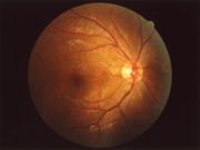
Fundus (eye)
The fundus of the eye is the interior surface of the eye, opposite the lens, and includes the retina, optic disc, macula, and posterior pole.[1] The fundus can be viewed with an ophthalmoscope.[1] The term may also be inclusive of Bruch's membrane and the choroid.
The eye's fundus is the only part of the human body where the microcirculation can be observed directly.[2] The diameter of the blood vessels around the optic disc is about 150 μm, and an ophthalmoscope allows observation of blood vessels with diameters as small as 10 μm.[2]
Diagnosis
Medical signs that can be detected from observation of eye fundus include haemorrhages, exudates, cotton wool spots, blood vessel abnormalities (tortuosity, pulsation and new vessels) and pigmentation.[3]
Physical Examination
Eyes
References
- ↑ 1.0 1.1 Cassin, B. and Solomon, S. Dictionary of Eye Terminology. Gainsville, Florida: Triad Publishing Company, 1990.
- ↑ 2.0 2.1 Ronald Pitts Crick, Peng Tee Khaw, "A Textbook of Clinical Ophthalmology: A Practical Guide to Disorders of the Eyes and Their Management", 3rd edition, World Scientific, 2003, ISBN 981-238-128-7
- ↑ Imran Akram, Adrian Rubinstein "Common retinal signs. An overview", "Optometry Today", 28/01/05, [1]
- ↑ http://picasaweb.google.com/mcmumbi/USMLEIIImages/
- ↑ http://picasaweb.google.com/mcmumbi/USMLEIIImages/
![Normal Fundus[4]](/images/a/ac/Normal_Fundus.jpg)

![Diabetic Fundus with hemorrhages[5]](/images/5/5b/DiabeticFundus%28notehemorrages%29.jpg)