File:Serous carcinoma cytology.jpg
Size of this preview: 800 × 533 pixels. Other resolutions: 2,560 × 1,707 pixels | 4,272 × 2,848 pixels.
Original file (4,272 × 2,848 pixels, file size: 870 KB, MIME type: image/jpeg)
Image courtesy of Wikipedia. Wikipedia (original file ‘’here’’). [http://wikipedia.org/licence Creative Commons BYSANC]
File history
Click on a date/time to view the file as it appeared at that time.
| Date/Time | Thumbnail | Dimensions | User | Comment | |
|---|---|---|---|---|---|
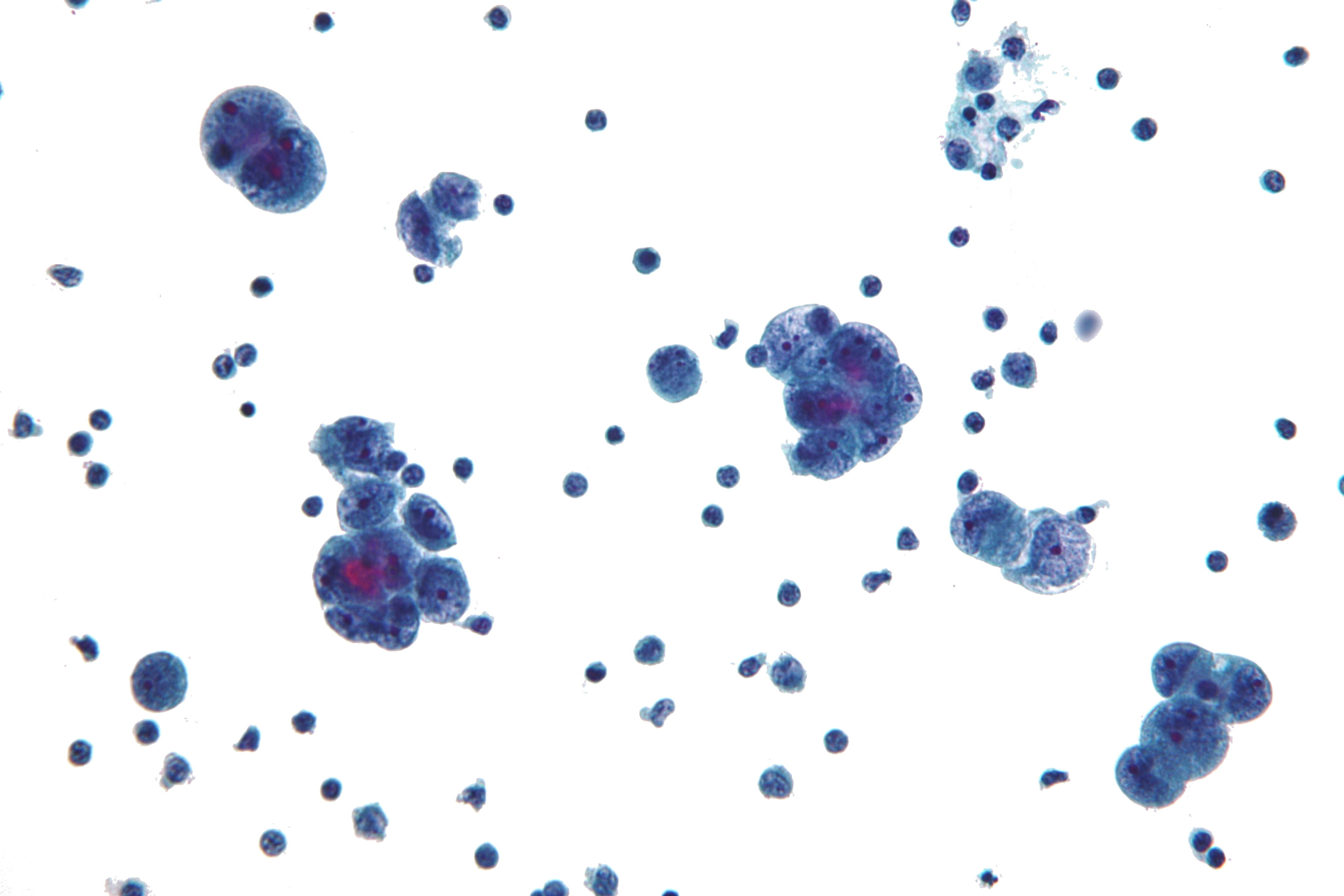
| current | 04:26, 13 April 2019 | | 4,272 × 2,848 (870 KB) | Ed Putiz (talk | contribs) | Specimen: Peritoneal Fluid. Microscopy showing seroous carcinoma with the following features: Marked intranuclear pleomorphism. Macronuclei. "Knobby" group borders (in large groups) - not apparent on this image. Hydropic vacuoles - not apparent on imag... |
| 21:38, 6 January 2016 |  | 1,024 × 683 (45 KB) | Ammu Susheela (talk | contribs) | Image courtesy of Wikipedia. [http://www.wikipedia.org Wikipedia] (original file [https://en.wikipedia.org/wiki/Primary_peritoneal_carcinoma#/media/File:Serous_carcinoma_cytology.jpg ‘’here’’]). [http://wikipedia.org/licence Creative... |
You cannot overwrite this file.
File usage
The following file is a duplicate of this file (more details):
.jpg)
The following page uses this file: