File:Papillary Tumor of the Pineal Region microscopic image 1.PNG: Difference between revisions
Jump to navigation
Jump to search
(Histology of the tumor showing papillary projections (a) based on hematoxylin eosin (H&E) staining. Immunohistochemistry demonstrating 20% positive Ki-67 (b), and the high expression of cytokeratin, an immunophenotype characteristic of PTPR (c).) |
(Sujit Routray uploaded a new version of File:Papillary Tumor of the Pineal Region microscopic image 1.PNG) |
(No difference)
| |
Latest revision as of 18:24, 24 November 2015
Histology of the tumor showing papillary projections (a) based on hematoxylin eosin (H&E) staining. Immunohistochemistry demonstrating 20% positive Ki-67 (b), and the high expression of cytokeratin, an immunophenotype characteristic of PTPR (c).
File history
Click on a date/time to view the file as it appeared at that time.
| Date/Time | Thumbnail | Dimensions | User | Comment | |
|---|---|---|---|---|---|
| current | 18:24, 24 November 2015 | 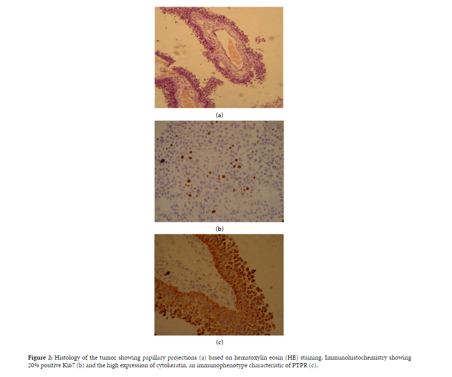 | 883 × 736 (390 KB) | Sujit Routray (talk | contribs) | Histology of the tumor showing papillary projections (a) based on hematoxylin eosin (H&E) staining. Immunohistochemistry demonstrating 20% positive Ki-67 (b), and the high expression of cytokeratin, an immunophenotype characteristic of PTPR (c). |
| 18:22, 24 November 2015 |  | 883 × 736 (390 KB) | Sujit Routray (talk | contribs) | Histology of the tumor showing papillary projections (a) based on hematoxylin eosin (H&E) staining. Immunohistochemistry demonstrating 20% positive Ki-67 (b), and the high expression of cytokeratin, an immunophenotype characteristic of PTPR (c). |
You cannot overwrite this file.
File usage
There are no pages that use this file.