File:Mast cell leukemia.jpg
Size of this preview: 600 × 600 pixels. Other resolution: 1,024 × 1,024 pixels.
Original file (1,024 × 1,024 pixels, file size: 99 KB, MIME type: image/jpeg)
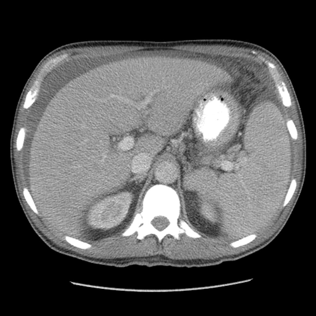
CT of the abdomen demonstrates ascites, hepatosplenomegaly and upper abdominal lymphadenopathy. Windowing to bone confirms the diffuse sclerosis seen on the plain films.
File history
Click on a date/time to view the file as it appeared at that time.
| Date/Time | Thumbnail | Dimensions | User | Comment | |
|---|---|---|---|---|---|
| current | 21:37, 1 December 2015 | | 1,024 × 1,024 (99 KB) | Nawal Muazam (talk | contribs) | CT of the abdomen demonstrates ascites, hepatosplenomegaly and upper abdominal lymphadenopathy. Windowing to bone confirms the diffuse sclerosis seen on the plain films. |
You cannot overwrite this file.
File usage
The following page uses this file: