File:Histoplasmosis02.jpeg
No higher resolution available.
Histoplasmosis02.jpeg (700 × 460 pixels, file size: 35 KB, MIME type: image/jpeg)
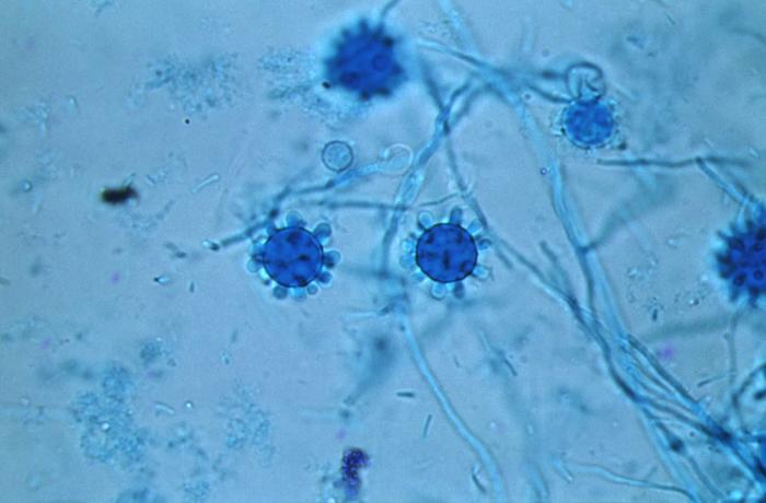
Under a magnification of 800X, this photomicrograph revealed some of the ultrastructural details exhibited by Histoplasma capsulatum fungal organisms that had been extracted from a Jamaican isolate, which included a number of tuberculate (knobby) spheroidal macroconidia, and diaphanous filamentous hyphae.
File history
Click on a date/time to view the file as it appeared at that time.
| Date/Time | Thumbnail | Dimensions | User | Comment | |
|---|---|---|---|---|---|
| current | 04:30, 12 December 2014 | | 700 × 460 (35 KB) | Jesus Hernandez (talk | contribs) | Under a magnification of 800X, this photomicrograph revealed some of the ultrastructural details exhibited by Histoplasma capsulatum fungal organisms that had been extracted from a Jamaican isolate, which included a number of tuberculate (knobby) spher... |
You cannot overwrite this file.
File usage
The following 2 pages use this file: