File:Coronavirus11.jpeg
Size of this preview: 606 × 599 pixels. Other resolution: 700 × 692 pixels.
Original file (700 × 692 pixels, file size: 217 KB, MIME type: image/jpeg)
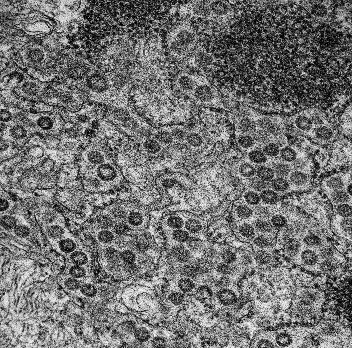
Produced by the National Institute of Allergy and Infectious Diseases (NIAID), this highly-magnified transmission electron micrograph (TEM) reveals the presence of numerous spherical-shaped Middle East Respiratory Syndrome Coronavirus (MERS-CoV) viral particles, which were found at the periphery of an infected MRC-5 cell (Human Fetal Lung Fibroblast).
File history
Click on a date/time to view the file as it appeared at that time.
| Date/Time | Thumbnail | Dimensions | User | Comment | |
|---|---|---|---|---|---|
| current | 20:41, 4 December 2014 | | 700 × 692 (217 KB) | Jesus Hernandez (talk | contribs) | Produced by the National Institute of Allergy and Infectious Diseases (NIAID), this highly-magnified transmission electron micrograph (TEM) reveals the presence of numerous spherical-shaped Middle East Respiratory Syndrome Coronavirus (MERS-CoV) viral ... |
You cannot overwrite this file.
File usage
The following page uses this file: