File:Chloroma histopathology micrograph (H&E stain).jpeg
Size of this preview: 800 × 533 pixels. Other resolution: 2,560 × 1,707 pixels.
Original file (2,560 × 1,707 pixels, file size: 1.08 MB, MIME type: image/jpeg)
Summary
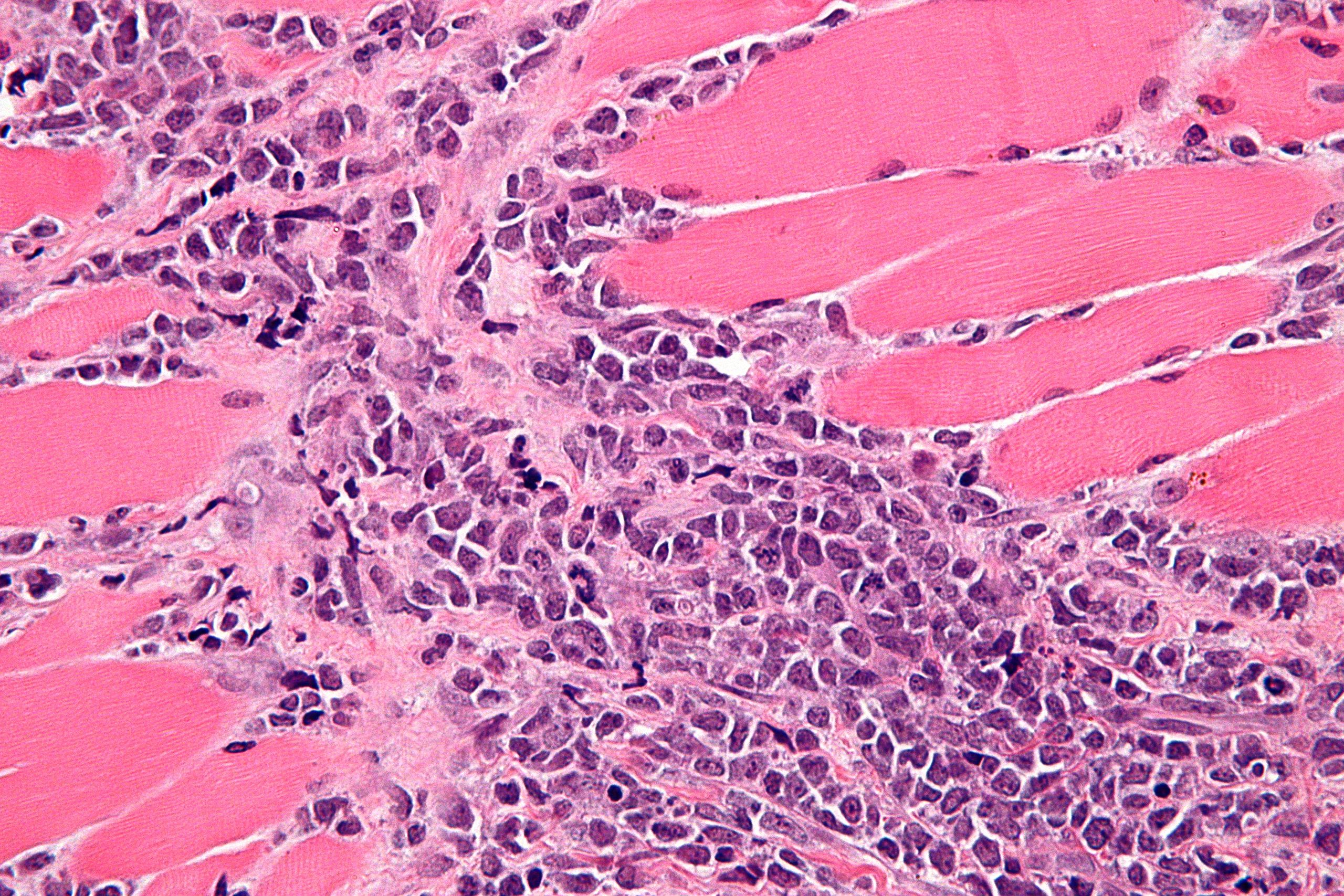
Histopathologic analysis of GS reveals many infiltrating myeloblasts. This can be seen in the course of AML. Courtesy of image from Wikipedia (By Nephron - Own work, CC BY-SA 3.0, https://commons.wikimedia.org/w/index.php?curid=15893726).
File history
Click on a date/time to view the file as it appeared at that time.
| Date/Time | Thumbnail | Dimensions | User | Comment | |
|---|---|---|---|---|---|
| current | 15:05, 2 May 2019 | | 2,560 × 1,707 (1.08 MB) | Parnian Jabbari (talk | contribs) | Histopathologic analysis of GS reveals many infiltrating myeloblasts. This can be seen in the course of AML. Courtesy of image from Wikipedia (By Nephron - Own work, CC BY-SA 3.0, https://commons.wikimedia.org/w/index.php?curid=15893726). |
You cannot overwrite this file.
File usage
The following page uses this file:
.jpeg&oldid=1566230)