The exact pathogenesis of elastofibroma dorsi is not fully understood. It is thought that elastofibroma dorsi is the result of subclinical microtrauma, reactive hyperplasia of elastic fibres and increased production of fibrous tissue. <ref name="pmid1417241">{{cite journal |vauthors=Machens HG, Mechtersheimer R, Göhring U, Schlag PN |title=Bilateral elastofibroma dorsi |journal=Ann. Thorac. Surg. |volume=54 |issue=4 |pages=774–6 |date=October 1992 |pmid=1417241 |doi= |url=}}</ref>
The exact pathogenesis of elastofibroma dorsi is not fully understood. It is thought that elastofibroma dorsi is the result of subclinical microtrauma, reactive hyperplasia of elastic fibres and increased production of fibrous tissue. <ref name="pmid1417241">{{cite journal |vauthors=Machens HG, Mechtersheimer R, Göhring U, Schlag PN |title=Bilateral elastofibroma dorsi |journal=Ann. Thorac. Surg. |volume=54 |issue=4 |pages=774–6 |date=October 1992 |pmid=1417241 |doi= |url=}}</ref>
Only 10% of patients has bilateral involvement. <ref name="pmid1503030">{{cite journal |vauthors=Kransdorf MJ, Meis JM, Montgomery E |title=Elastofibroma: MR and CT appearance with radiologic-pathologic correlation |journal=AJR Am J Roentgenol |volume=159 |issue=3 |pages=575–9 |date=September 1992 |pmid=1503030 |doi=10.2214/ajr.159.3.1503030 |url=}}</ref>
OR
OR
Line 78:
Line 80:
==Causes==
==Causes==
Disease name] may be caused by [cause1], [cause2], or [cause3].
Disease name] may be caused by [cause1], [cause2], or [cause3].
The cause of elastofibroma dorsi has not been identified.
OR
OR
Line 89:
Line 93:
OR
OR
The cause of [disease name] has not been identified. To review risk factors for the development of [disease name], click [[Pericarditis causes#Overview|here]].
The cause of elastofibroma dorsi has not been identified. To review risk factors for the development of [disease name], click [[Pericarditis causes#Overview|here]].
==Differentiating ((Page name)) from Other Diseases==
[Disease name] must be differentiated from other diseases that cause [clinical feature 1], [clinical feature 2], and [clinical feature 3], such as [differential dx1], [differential dx2], and [differential dx3].
OR
==Differentiating Elastofibroma dorsi from Other Diseases==
[Disease name] must be differentiated from [[differential dx1], [differential dx2], and [differential dx3].
Elastofibroma dorsi must be differentiated from desmoid tumours, neurofibroma and liposarcoma. <ref name="pmid185512042">{{cite journal |vauthors=Tetikkurt C, Tetikkurt S, Bayar N |title=Diagnosis of elastofibroma |journal=Can. Respir. J. |volume=15 |issue=4 |pages=217–8 |date=2008 |pmid=18551204 |pmc=2677955 |doi=10.1155/2008/638624 |url=}}</ref>
==Epidemiology and Demographics==
==Epidemiology and Demographics==
The incidence/prevalence of [disease name] is approximately [number range] per 100,000 individuals worldwide.
The incidence/prevalence of [disease name] is approximately [number range] per 100,000 individuals worldwide.
but an autopsy series found a frequency of 11.2% in men and 24.4% in women
OR
OR
Line 207:
Line 210:
===History and Symptoms===
===History and Symptoms===
The majority of patients with [disease name] are asymptomatic.
The majority of patients with elastofibroma dorsi are asymptomatic. Elastofibroma may present with:<ref name="pmid2652048">{{cite journal |vauthors=Greenberg JA, Lockwood RC |title=Elastofibroma dorsi. A case report and review of the literature |journal=Orthop Rev |volume=18 |issue=3 |pages=329–33 |date=March 1989 |pmid=2652048 |doi= |url=}}</ref>
* Painless swelling
OR
* Pain ( less than 10% of patients)
The hallmark of [disease name] is [finding]. A positive history of [finding 1] and [finding 2] is suggestive of [disease name]. The most common symptoms of [disease name] include [symptom 1], [symptom 2], and [symptom 3]. Common symptoms of [disease] include [symptom 1], [symptom 2], and [symptom 3]. Less common symptoms of [disease name] include [symptom 1], [symptom 2], and [symptom 3].
The hallmark of [disease name] is [finding]. A positive history of [finding 1] and [finding 2] is suggestive of [disease name]. The most common symptoms of [disease name] include [symptom 1], [symptom 2], and [symptom 3]. Common symptoms of [disease] include [symptom 1], [symptom 2], and [symptom 3]. Less common symptoms of [disease name] include [symptom 1], [symptom 2], and [symptom 3].
Line 255:
Line 258:
===X-ray===
===X-ray===
There are no x-ray findings associated with [disease name].
OR
An x-ray may be helpful in the diagnosis of [disease name]. Findings on an x-ray suggestive of/diagnostic of [disease name] include [finding 1], [finding 2], and [finding 3].
OR
There are no x-ray findings associated with [disease name]. However, an x-ray may be helpful in the diagnosis of complications of [disease name], which include [complication 1], [complication 2], and [complication 3].
An x-ray may be helpful in the diagnosis of elastofibroma. Findings on an x-ray suggestive of elastofibroma include soft tissue density in the periscapular region. X-ray may be normal. <ref name="pmid18551204">{{cite journal |vauthors=Tetikkurt C, Tetikkurt S, Bayar N |title=Diagnosis of elastofibroma |journal=Can. Respir. J. |volume=15 |issue=4 |pages=217–8 |date=2008 |pmid=18551204 |pmc=2677955 |doi=10.1155/2008/638624 |url=}}</ref>
===Echocardiography or Ultrasound===
===Echocardiography or Ultrasound===
Line 277:
Line 273:
===CT scan===
===CT scan===
There are no CT scan findings associated with [disease name].
CT scan may be helpful in the diagnosis of elastofibroma dorsi. Findings on CT scan suggestive of elastofibroma dorsi include a heterogenous soft tissue mass with poorly defined margins. <ref name="pmid8998883">{{cite journal |vauthors=Hoffman JK, Klein MH, McInerney VK |title=Bilateral elastofibroma: a case report and review of the literature |journal=Clin. Orthop. Relat. Res. |volume= |issue=325 |pages=245–50 |date=April 1996 |pmid=8998883 |doi= |url=}}</ref>
OR
[Location] CT scan may be helpful in the diagnosis of [disease name]. Findings on CT scan suggestive of/diagnostic of [disease name] include [finding 1], [finding 2], and [finding 3].
OR
There are no CT scan findings associated with [disease name]. However, a CT scan may be helpful in the diagnosis of complications of [disease name], which include [complication 1], [complication 2], and [complication 3].
===MRI===
===MRI===
There are no MRI findings associated with [disease name].
Magnetic resonance imaging is the most useful diagnostic tool for diagnosis of elastofibroma dorsi. <ref name="pmid14648789">{{cite journal |vauthors=Domanski HA, Carlén B, Sloth M, Rydholm A |title=Elastofibroma dorsi has distinct cytomorphologic features, making diagnostic surgical biopsy unnecessary: cytomorphologic study with clinical, radiologic, and electron microscopic correlations |journal=Diagn. Cytopathol. |volume=29 |issue=6 |pages=327–33 |date=December 2003 |pmid=14648789 |doi=10.1002/dc.10381 |url=}}</ref>
OR
OR
[Location] MRI may be helpful in the diagnosis of [disease name]. Findings on MRI suggestive of/diagnostic of [disease name] include [finding 1], [finding 2], and [finding 3].
[Location] MRI may be helpful in the Magnetic resonance imaging is the most useful diagnostic tool [disease name]. Findings on MRI suggestive of/diagnostic of [disease name] include [finding 1], [finding 2], and [finding 3].
OR
OR
Line 299:
Line 287:
===Other Imaging Findings===
===Other Imaging Findings===
There are no other imaging findings associated with [disease name].
OR
Positron emission tomography/computed tomography (PET/CT) may be helpful in the diagnosis of elastofibroma. Findings on PET/CT suggestive of elastofibroma include low to moderate metabolic activity in these patients. PET/CT shows low-grade diffuse 18F fluorodeoxyglucose uptake. <ref name="pmid16100483">{{cite journal |vauthors=Patrikeos A, Breidahl W, Robins P |title=F-18 FDG uptake associated with Elastofibroma dorsi |journal=Clin Nucl Med |volume=30 |issue=9 |pages=617–8 |date=September 2005 |pmid=16100483 |doi= |url=}}</ref>
[Imaging modality] may be helpful in the diagnosis of [disease name]. Findings on an [imaging modality] suggestive of/diagnostic of [disease name] include [finding 1], [finding 2], and [finding 3].
===Other Diagnostic Studies===
===Other Diagnostic Studies===
There are no other diagnostic studies associated with [disease name].
OR
[Diagnostic study] may be helpful in the diagnosis of [disease name]. Findings suggestive of/diagnostic of [disease name] include [finding 1], [finding 2], and [finding 3].
Needle aspiration biopsy is helpful in the diagnosis of elastofibroma and exclude sarcoma. Findings suggestive of/diagnostic of [disease name] include [finding 1], [finding 2], and [finding 3].
Elastofibroma was first discovered by Jarvi and Saxen, in 1961.[1]
The association between [important risk factor/cause] and [disease name] was made in/during [year/event].
In [year], [scientist] was the first to discover the association between [risk factor] and the development of [disease name].
In [year], [gene] mutations were first implicated in the pathogenesis of [disease name].
There have been several outbreaks of [disease name], including -----.
In [year], [diagnostic test/therapy] was developed by [scientist] to treat/diagnose [disease name].
Classification
There is no established system for the classification of [disease name].
OR
[Disease name] may be classified according to [classification method] into [number] subtypes/groups: [group1], [group2], [group3], and [group4].
OR
[Disease name] may be classified into [large number > 6] subtypes based on [classification method 1], [classification method 2], and [classification method 3].
[Disease name] may be classified into several subtypes based on [classification method 1], [classification method 2], and [classification method 3].
OR
Based on the duration of symptoms, [disease name] may be classified as either acute or chronic.
OR
If the staging system involves specific and characteristic findings and features:
According to the [staging system + reference], there are [number] stages of [malignancy name] based on the [finding1], [finding2], and [finding3]. Each stage is assigned a [letter/number1] and a [letter/number2] that designate the [feature1] and [feature2].
OR
The staging of [malignancy name] is based on the [staging system].
OR
There is no established system for the staging of [malignancy name].
Pathophysiology
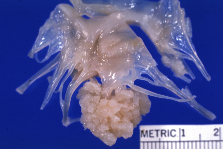
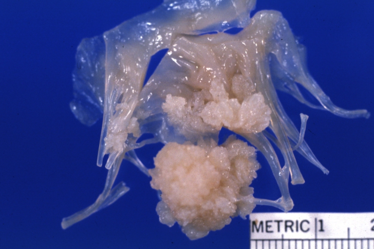
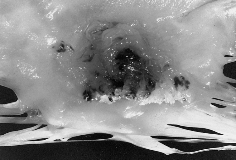
Elastofibroma dorsi is a rare, slow growing, ill-defined soft tissue mass of the chest wall. It occurs most in the periscapular region.It is commonly located beneath latissimus dorsi and rhomboid major muscles near to the inferior angle of the scapula. It is a benign neoplasm with clinical appearence of a malignant tumor. [2]
The exact pathogenesis of elastofibroma dorsi is not fully understood. It is thought that elastofibroma dorsi is the result of subclinical microtrauma, reactive hyperplasia of elastic fibres and increased production of fibrous tissue. [3]
Only 10% of patients has bilateral involvement. [4]
OR
[Pathogen name] is usually transmitted via the [transmission route] route to the human host.
OR
Following transmission/ingestion, the [pathogen] uses the [entry site] to invade the [cell name] cell.
OR
[Disease or malignancy name] arises from [cell name]s, which are [cell type] cells that are normally involved in [function of cells].
OR
The progression to [disease name] usually involves the [molecular pathway].
OR
The pathophysiology of [disease/malignancy] depends on the histological subtype.
Causes
Disease name] may be caused by [cause1], [cause2], or [cause3].
The cause of elastofibroma dorsi has not been identified.
OR
Common causes of [disease] include [cause1], [cause2], and [cause3].
OR
The most common cause of [disease name] is [cause 1]. Less common causes of [disease name] include [cause 2], [cause 3], and [cause 4].
OR
The cause of elastofibroma dorsi has not been identified. To review risk factors for the development of [disease name], click here.
Differentiating Elastofibroma dorsi from Other Diseases
Elastofibroma dorsi must be differentiated from desmoid tumours, neurofibroma and liposarcoma. [5]
Epidemiology and Demographics
The incidence/prevalence of [disease name] is approximately [number range] per 100,000 individuals worldwide.
but an autopsy series found a frequency of 11.2% in men and 24.4% in women
OR
In [year], the incidence/prevalence of [disease name] was estimated to be [number range] cases per 100,000 individuals worldwide.
OR
In [year], the incidence of [disease name] is approximately [number range] per 100,000 individuals with a case-fatality rate of [number range]%.
Patients of all age groups may develop [disease name].
OR
The incidence of [disease name] increases with age; the median age at diagnosis is [#] years.
OR
[Disease name] commonly affects individuals younger than/older than [number of years] years of age.
OR
[Chronic disease name] is usually first diagnosed among [age group].
There is no racial predilection to [disease name].
OR
[Disease name] usually affects individuals of the [race 1] race. [Race 2] individuals are less likely to develop [disease name].
[Disease name] affects men and women equally.
OR
[Gender 1] are more commonly affected by [disease name] than [gender 2]. The [gender 1] to [gender 2] ratio is approximately [number > 1] to 1.
The majority of [disease name] cases are reported in [geographical region].
OR
[Disease name] is a common/rare disease that tends to affect [patient population 1] and [patient population 2].
Risk Factors
There are no established risk factors for [disease name].
OR
The most potent risk factor in the development of [disease name] is [risk factor 1]. Other risk factors include [risk factor 2], [risk factor 3], and [risk factor 4].
OR
Common risk factors in the development of [disease name] include [risk factor 1], [risk factor 2], [risk factor 3], and [risk factor 4].
OR
Common risk factors in the development of [disease name] may be occupational, environmental, genetic, and viral.
Screening
There is insufficient evidence to recommend routine screening for [disease/malignancy].
OR
According to the [guideline name], screening for [disease name] is not recommended.
OR
According to the [guideline name], screening for [disease name] by [test 1] is recommended every [duration] among patients with [condition 1], [condition 2], and [condition 3].
Natural History, Complications, and Prognosis
If left untreated, [#]% of patients with [disease name] may progress to develop [manifestation 1], [manifestation 2], and [manifestation 3].
OR
Common complications of [disease name] include [complication 1], [complication 2], and [complication 3].
OR
Prognosis is generally excellent/good/poor, and the 1/5/10-year mortality/survival rate of patients with [disease name] is approximately [#]%.
Diagnosis
Diagnostic Study of Choice
The diagnosis of [disease name] is made when at least [number] of the following [number] diagnostic criteria are met: [criterion 1], [criterion 2], [criterion 3], and [criterion 4].
OR
The diagnosis of [disease name] is based on the [criteria name] criteria, which include [criterion 1], [criterion 2], and [criterion 3].
OR
The diagnosis of [disease name] is based on the [definition name] definition, which includes [criterion 1], [criterion 2], and [criterion 3].
OR
There are no established criteria for the diagnosis of [disease name].
History and Symptoms
The majority of patients with elastofibroma dorsi are asymptomatic. Elastofibroma may present with:[6]
Painless swelling
Pain ( less than 10% of patients)
The hallmark of [disease name] is [finding]. A positive history of [finding 1] and [finding 2] is suggestive of [disease name]. The most common symptoms of [disease name] include [symptom 1], [symptom 2], and [symptom 3]. Common symptoms of [disease] include [symptom 1], [symptom 2], and [symptom 3]. Less common symptoms of [disease name] include [symptom 1], [symptom 2], and [symptom 3].
Physical Examination
Patients with [disease name] usually appear [general appearance]. Physical examination of patients with [disease name] is usually remarkable for [finding 1], [finding 2], and [finding 3].
OR
Common physical examination findings of [disease name] include [finding 1], [finding 2], and [finding 3].
OR
The presence of [finding(s)] on physical examination is diagnostic of [disease name].
OR
The presence of [finding(s)] on physical examination is highly suggestive of [disease name].
Laboratory Findings
An elevated/reduced concentration of serum/blood/urinary/CSF/other [lab test] is diagnostic of [disease name].
OR
Laboratory findings consistent with the diagnosis of [disease name] include [abnormal test 1], [abnormal test 2], and [abnormal test 3].
OR
[Test] is usually normal among patients with [disease name].
OR
Some patients with [disease name] may have elevated/reduced concentration of [test], which is usually suggestive of [progression/complication].
OR
There are no diagnostic laboratory findings associated with [disease name].
Electrocardiogram
There are no ECG findings associated with [disease name].
OR
An ECG may be helpful in the diagnosis of [disease name]. Findings on an ECG suggestive of/diagnostic of [disease name] include [finding 1], [finding 2], and [finding 3].
X-ray
An x-ray may be helpful in the diagnosis of elastofibroma. Findings on an x-ray suggestive of elastofibroma include soft tissue density in the periscapular region. X-ray may be normal. [7]
Echocardiography or Ultrasound
There are no echocardiography/ultrasound findings associated with [disease name].
OR
Echocardiography/ultrasound may be helpful in the diagnosis of [disease name]. Findings on an echocardiography/ultrasound suggestive of/diagnostic of [disease name] include [finding 1], [finding 2], and [finding 3].
OR
There are no echocardiography/ultrasound findings associated with [disease name]. However, an echocardiography/ultrasound may be helpful in the diagnosis of complications of [disease name], which include [complication 1], [complication 2], and [complication 3].
CT scan
CT scan may be helpful in the diagnosis of elastofibroma dorsi. Findings on CT scan suggestive of elastofibroma dorsi include a heterogenous soft tissue mass with poorly defined margins. [8]
MRI
Magnetic resonance imaging is the most useful diagnostic tool for diagnosis of elastofibroma dorsi. [9]
OR
[Location] MRI may be helpful in the Magnetic resonance imaging is the most useful diagnostic tool [disease name]. Findings on MRI suggestive of/diagnostic of [disease name] include [finding 1], [finding 2], and [finding 3].
OR
There are no MRI findings associated with [disease name]. However, a MRI may be helpful in the diagnosis of complications of [disease name], which include [complication 1], [complication 2], and [complication 3].
Other Imaging Findings
Positron emission tomography/computed tomography (PET/CT) may be helpful in the diagnosis of elastofibroma. Findings on PET/CT suggestive of elastofibroma include low to moderate metabolic activity in these patients. PET/CT shows low-grade diffuse 18F fluorodeoxyglucose uptake. [10]
Other Diagnostic Studies
Needle aspiration biopsy is helpful in the diagnosis of elastofibroma and exclude sarcoma. Findings suggestive of/diagnostic of [disease name] include [finding 1], [finding 2], and [finding 3].
OR
Other diagnostic studies for [disease name] include [diagnostic study 1], which demonstrates [finding 1], [finding 2], and [finding 3], and [diagnostic study 2], which demonstrates [finding 1], [finding 2], and [finding 3].
Treatment
Medical Therapy
There is no treatment for [disease name]; the mainstay of therapy is supportive care.
OR
Supportive therapy for [disease name] includes [therapy 1], [therapy 2], and [therapy 3].
OR
The majority of cases of [disease name] are self-limited and require only supportive care.
OR
[Disease name] is a medical emergency and requires prompt treatment.
OR
The mainstay of treatment for [disease name] is [therapy].
OR
The optimal therapy for [malignancy name] depends on the stage at diagnosis.
OR
[Therapy] is recommended among all patients who develop [disease name].
OR
Pharmacologic medical therapy is recommended among patients with [disease subclass 1], [disease subclass 2], and [disease subclass 3].
OR
Pharmacologic medical therapies for [disease name] include (either) [therapy 1], [therapy 2], and/or [therapy 3].
OR
Empiric therapy for [disease name] depends on [disease factor 1] and [disease factor 2].
OR
Patients with [disease subclass 1] are treated with [therapy 1], whereas patients with [disease subclass 2] are treated with [therapy 2].
Surgery
Surgical intervention is not recommended for the management of [disease name].
OR
Surgery is not the first-line treatment option for patients with [disease name]. Surgery is usually reserved for patients with either [indication 1], [indication 2], and [indication 3]
OR
The mainstay of treatment for [disease name] is medical therapy. Surgery is usually reserved for patients with either [indication 1], [indication 2], and/or [indication 3].
OR
The feasibility of surgery depends on the stage of [malignancy] at diagnosis.
OR
Surgery is the mainstay of treatment for [disease or malignancy].
Primary Prevention
There are no established measures for the primary prevention of [disease name].
OR
There are no available vaccines against [disease name].
OR
Effective measures for the primary prevention of [disease name] include [measure1], [measure2], and [measure3].
OR
[Vaccine name] vaccine is recommended for [patient population] to prevent [disease name]. Other primary prevention strategies include [strategy 1], [strategy 2], and [strategy 3].
Secondary Prevention
There are no established measures for the secondary prevention of [disease name].
OR
Effective measures for the secondary prevention of [disease name] include [strategy 1], [strategy 2], and [strategy 3].
References
↑JARVI O, SAXEN E (1961). "Elastofibroma dorse". Acta Pathol Microbiol Scand Suppl. 51(Suppl 144): 83–4. PMID13789598.
↑Freixinet J, Rodríguez P, Hussein M, Sanromán B, Herrero J, Gil R (August 2008). "Elastofibroma of the thoracic wall". Interact Cardiovasc Thorac Surg. 7 (4): 626–8. doi:10.1510/icvts.2007.174722. PMID18407963.
↑Machens HG, Mechtersheimer R, Göhring U, Schlag PN (October 1992). "Bilateral elastofibroma dorsi". Ann. Thorac. Surg. 54 (4): 774–6. PMID1417241.
↑Kransdorf MJ, Meis JM, Montgomery E (September 1992). "Elastofibroma: MR and CT appearance with radiologic-pathologic correlation". AJR Am J Roentgenol. 159 (3): 575–9. doi:10.2214/ajr.159.3.1503030. PMID1503030.
↑Hoffman JK, Klein MH, McInerney VK (April 1996). "Bilateral elastofibroma: a case report and review of the literature". Clin. Orthop. Relat. Res. (325): 245–50. PMID8998883.
↑Domanski HA, Carlén B, Sloth M, Rydholm A (December 2003). "Elastofibroma dorsi has distinct cytomorphologic features, making diagnostic surgical biopsy unnecessary: cytomorphologic study with clinical, radiologic, and electron microscopic correlations". Diagn. Cytopathol. 29 (6): 327–33. doi:10.1002/dc.10381. PMID14648789.
↑Patrikeos A, Breidahl W, Robins P (September 2005). "F-18 FDG uptake associated with Elastofibroma dorsi". Clin Nucl Med. 30 (9): 617–8. PMID16100483.
Elastofibroma is an ill-defined fibroelastic tumor-like condition made up of enlarged and irregular elastic fibers. On gross pathology, ill defined, nonencapsulated, rubbery, and firm, white lesion with interspersed fat are characteristic findings of elastofibroma. On microscopic histopathological analysis, heavy dense bands of collagenous tissue dissected by fat and abnormal elastic fibers are characteristic findings of elastofibroma . The elastic fibers are usually quite large and are easily identified. The elastic fibers are coarse, thick, and darkly eosinophilic, often fragmented into globules, creating a "string of pearls" or "pipe cleaner" appearance. Degeneration will cause the elastic fibers to appear as globules with a serrated or prickled edge. Elastofibroma may be caused by either trauma, genetic mutation, or systemic enzyme defects. Elastofibroma must be differentiated from other diseases that cause soft tissue tumor such as: spindle cell lipoma, nuchal-type fibroma, and fibromatosis colli. Elastofibroma may also be diagnosed using biopsy and histochemistry. Surgery is the mainstay of therapy for elastofibroma.
Pathophysiology
Elastofibroma, also called elastofibroma dorsi, is an ill-defined fibroelastic tumor-like condition made up of enlarged and irregular elastic fibers. [1][2]
The tumor develops very specifically in the subscapular or infrascapular area, deep to the muscle, and can be attached to periosteum of ribs. It is usually between the shoulder blade and the lower neck, with rare tumors reported in the chest wall. [1][3][2]
The genetic mutation in has been associated alterations of short arm of chromosome 1 with the development of elastofibroma.
On gross pathology, ill defined, nonencapsulated, rubbery, and firm, white lesion with interspersed fat are characteristic findings of elastofibroma.
On microscopic histopathological analysis, heavy dense bands of collagenous tissue dissected by fat and abnormal elastic fibers are characteristic findings of elastofibroma. The elastic fibers are often quite large and are easily identified. The elastic fibers are coarse, thick, and darkly eosinophilic, often fragmented into globules, creating a "string of pearls" or "pipe cleaner" appearance. Degeneration will cause the elastic fibers to appear as globules with a serrated or prickled edge.
Elastofibroma
Elastofibroma
Papillary Fibroelastoma: When located on the mitral valve, these tumors are usually on the anterior leaflet of the atrial surface.
Causes
Elastofibroma may be caused by either trauma, genetic mutation, or systemic enzyme defects.
Differentiating Elastofibroma from other Diseases
Elastofibroma must be differentiated from other diseases that cause soft tissue tumor such as:
↑ 2.02.1Briccoli, A.; Casadei, R.; Di Renzo, M.; Favale, L.; Bacchini, P.; Bertoni, F. (2000). "Elastofibroma dorsi". Surgery today. 30 (2): 147–152. doi:10.1007/pl00010063. PMID10664338.
↑Mortman, K. D.; Hochheiser, G. M.; Giblin, E. M.; Manon-Matos, Y.; Frankel, K. M. (2007). "Elastofibroma Dorsi: Clinicopathologic Review of 6 Cases". The Annals of Thoracic Surgery. 83 (5): 1894–1897. doi:10.1016/j.athoracsur.2006.11.050. PMID17462431.