Dextrocardia electrocardiogram
|
Dextrocardia Microchapters |
|
Diagnosis |
|---|
|
Treatment |
|
Dextrocardia electrocardiogram On the Web |
|
American Roentgen Ray Society Images of Dextrocardia electrocardiogram |
|
Risk calculators and risk factors for Dextrocardia electrocardiogram |
Editor-In-Chief: C. Michael Gibson, M.S., M.D. [1]; Associate Editors-In-Chief: Priyamvada Singh, M.B.B.S. [[2]]; Cafer Zorkun, M.D., Ph.D. [3]; Keri Shafer, M.D. [4]; Claudia Hochberg, M.D.; Assistant Editor-In-Chief: Kristin Feeney, B.S. [[5]]
Overview
Electrocardiogram may be used as a diagnostic tool in the evaluation of an atrial septal defect. ECG findings associated with dextrocardia include an R wave inversion.
Electrocardiogram
It is possible to distinguish lead reversal and dextrocardia by watching the precordial leads. Dextrocardia will show an R wave inversion, whereas lead reversal will not.
The bottom EKG shows marked right axis deviation and loss of voltage across the precordium. There are also inverted P waves in leads I and aVL. The differential for inverted P waves in lead I and aVL is Dextrocardia vs Reversed Arm Leads. Since there is loss of voltage across the precordium this is Dextrocardia.
Special considerations
ECG leads must be placed in reversed positions on a person with Dextrocardia.
- Shown below is am image of EKG in a a patient with Dextrocardia .
EKG demonstrates right axis deviation and loss of voltage across the precordium. There are also inverted p waves in leads I and aVL.

-
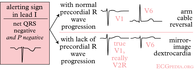
Right and left arm lead reversal can be distinguished from the (much rarer) dextrocardia by looking at the precordial R wave progression.
-
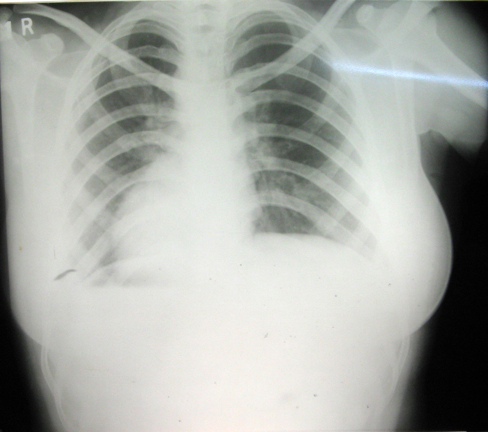
EKG in a patient with dextrocardia. This EKG shows marked right axis deviation and loss of voltage across the precordium. There are also inverted p waves in leads I and aVL. The differential for inverted p waves in lead I and aVL is Dextrocardia or Reversed Arm Leads. Since there is loss of voltage across the precordium this is Dextrocardia.
