Dextrocardia: Difference between revisions

Jump to navigation Jump to search
No edit summary
No edit summary
Line 1: Line 1:
'''For the WikiPatient page for this topic, click [[Dextrocardia (patient information)|here]]'''
{{CMG}}; '''Associate Editors-In-Chief:''' [[Priyamvada Singh|Priyamvada Singh, MBBS]] [[mailto:psingh@perfuse.org]]; {{CZ}}; [[User:KeriShafer|Keri Shafer, M.D.]] [mailto:kshafer@bidmc.harvard.edu];  Claudia Hochberg, M.D.; '''Assistant Editor-In-Chief:''' [[Kristin Feeney|Kristin Feeney, B.S.]] [[mailto:kfeeney@perfuse.org]]
{{Infobox_Disease |
{{Infobox_Disease |
   Name          = Dextrocardia |
   Name          = Dextrocardia |
Line 18: Line 15:
   MeshNumber    = C14.240.400.280 |
   MeshNumber    = C14.240.400.280 |
}}
}}
==Overview==
{{Dextrocardia}}
'''Dextrocardia''' refers to the heart being situated on the right side of the bodyIf all visceral organs are mirror reversed, the correct term is Dextrocardia Situs Inversus Totalis. Individuals with both dextrocardia and situs inversus have a lower incidence of congenital heart defects than do people with only one of the two conditions.
{{CMG}}; '''Associate Editors-In-Chief:''' [[Priyamvada Singh|Priyamvada Singh, MBBS]] [[mailto:psingh@perfuse.org]]; {{CZ}}; [[User:KeriShafer|Keri Shafer, M.D.]] [mailto:kshafer@bidmc.harvard.edu]; Claudia Hochberg, M.D.; '''Assistant Editor-In-Chief:''' [[Kristin Feeney|Kristin Feeney, B.S.]] [[mailto:kfeeney@perfuse.org]]


==History==
==[[Type page name here overview|Overview]]==
Dextorocardia was decribed by Fabricious in 1606. Situs inversus with dextrocardia (totalis) was first described by Severinus in 1643.


==Epidemiology==
==[[Type page name here pathophysiology|Pathophysiology]]==
Dextrocardia is believed to occur in approximately 1 in a 100 people, while 1 in 1,000 of these will have [[Situs Inversus]].


[[Kartagener’s syndrome]] occurs in approximately 1 in 15-25 of patients with Totalis (situs inversus with dextrocardia). Kartagener Syndrome (KS), is a rare autosomal recessive genetic disorder caused by a defect in the action of the tiny hairs (cilia) lining the respiratory tract. Specifically, it is a defect in a gene coding for left-right dynein (lrd), a key structural protein in cilia.<ref>Cleveland JC Jr, Shroyer AL, Chen AY, et al: Off-pump coronary artery bypass grafting decreases risk-adjusted mortality and morbidity. Ann Thorac Surg 2001;72: 1282-1289.</ref>
==[[Type page name here epidemiology and demographics|Epidemiology and demographics]]==


==Pathophysiology==
==[[Type page name here natural history|Natural history, Complications, and Prognosis]]==
Dextrocardia is not associated with any medical sequela.


==Diagnosis==
==[[Type page name here causes|Causes]]==
===Electrocardiogram===


It is possible to distinguish lead reversal and [[dextrocardia]] by watching the precordial leads. Dextrocardia will show an R wave inversion, whereas lead reversal will not.
==[[Type page name here differential diagnosis|Differentiating Type page name here from other Disorders]]==
The bottom EKG shows marked [[right axis deviation]] and loss of voltage across the precordium. There are also inverted [[P wave]]s in leads I and aVL. The differential for inverted P waves in lead I and aVL is Dextrocardia vs Reversed Arm Leads. Since there is loss of voltage across the precordium this is Dextrocardia.


<div align="left">
==Diagnosis==
<gallery heights="175" widths="175">
[[Type page name here history and symptoms|History and Symptoms]] | [[Type page name here physical examination|Physical Examination]] | [[Type page name here laboratory tests|Laboratory Tests]] | [[Type page name here electrocardiogram | Electrocardiogram]] | [[Type page name here chest x ray|Chest X Ray]] | [[Type page name here MRI|MRI]] | [[Type page name here CT|CT]] | [[Type page name here echocardiography|Echocardiography]] | [[Type page name here other imaging findings|Other Imaging Findings]] | [[Type page name here other diagnostic studies|Other Diagnostic Studies]]
Image:cableReversal1.png|Right and left arm lead reversal can be distinguished from the (much rarer) dextrocardia by looking at the precordial R wave progression.
Image:Dextrocardia.jpg|EKG in a patient with dextrocardia. This EKG shows marked right axis deviation and loss of voltage across the precordium. There are also inverted p waves in leads I and aVL. The differential for inverted p waves in lead I and aVL is Dextrocardia or Reversed Arm Leads. Since there is loss of voltage across the precordium this is Dextrocardia.
</gallery>
</div>


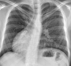
===Chest X Ray===
==Treatment==
'''Medical:''' [[Type page name here medical therapy|Medical Therapy]]


Shown to the right is a chest X ray in a patient with Dextrocardia. Note that the heart is on the right side of the chest.
'''Surgical:''' [[Type page name here surgery|Surgery]]


<div align="left">
[[Type page name here primary prevention|Primary Prevention]] | [[Type page name here secondary prevention|Secondary Prevention]] | [[Type page name here cost-effectiveness of therapy|Cost-Effectiveness of Therapy]] | [[Type page name here future or investigational therapies|Future or Investigational Therapies]]
<gallery heights="175" widths="175">
Image:Dextrocardia cxr.jpg|
</gallery>
</div>


===Coronary Angiography===
* Dextrocardia Angiogram 1
<youtube v=r570-FIuVCc/>
* Dextrocardia Angiogram 2
<youtube v=Kd1LjLJWvTQ/>
* Dextrocardia Angiogram 3
<youtube v=rIQqCAgLk1Y/>
===Fetal and Postnatal Diagnosis by Ultrasound, Magnetic Resonance Imaging (MRI), and Chest X Ray===
The presenting twin of this 22 week gestation has dextrocardia. Images supported by NIH grant NS37945
<div align="left">
<gallery heights="175" widths="175">
Image:Dextrocardia prenatal ultrasound.jpg
Image:Dextrocardia mri1.jpg
Image:Dextrocardia mri2.jpg
</gallery>
</div>
<div align="left">
<gallery heights="175" widths="175">
Image:Dextrocardia mri3.jpg
Image:Dextrocardia postnatal.jpg
</gallery>
</div>
===Performing an EKG===
ECG leads must be placed in reversed positions on a person with Dextrocardia.
===Defibrillating a Patient with Dextrocardia===
When defibrillating a patient with dextrocardia, the pads should be placed in reverse positions. That is, instead of upper right and lower left, pads should be placed upper left and lower right.
==Treatment==
===Coronary Artery Bypass Grafting===
CABG can be performed in the patient with dextrocardia. A right [[internal mammary artery]] may be used as a conduit to the [[left anterior descending artery]].<ref>Irvin RG, Ballenger JF: Coronary artery bypass in a patient with situs inversus. Chest 1982;81:380-381.</ref> <ref>Mesa JM, Aroca A, Frutos A, et al: Situs inversus and myocardial revascularization: Case report. J Cardiovasc Surg 1995;36: 571-572.</ref> <ref>Cleveland JC Jr, Shroyer AL, Chen AY, et al: Off-pump coronary artery bypass grafting decreases risk-adjusted mortality and morbidity. Ann Thorac Surg 2001;72: 1282-1289.</ref>


==References==
==References==
Line 101: Line 45:


==Additional resources==
* [http://www.rch.org.au/cardiology/health-info.cfm?doc_id=3538 Overview at rch.org.au]


{{Congenital malformations and deformations of circulatory system}}
{{SIB}}


[[Category:Cardiology]]
[[Category:Cardiology]]

Revision as of 12:53, 10 August 2011

For the WikiPatient page for this topic, click here

Dextrocardia
ICD-10 Q24.0
ICD-9 746.87
DiseasesDB 3617
MeSH C14.240.400.280

Dextrocardia Microchapters

Home

Patient Information

Overview

Historical Perspective

Classification

Pathophysiology

Causes

Differentiating Dextrocardia from other Diseases

Epidemiology and Demographics

Risk Factors

Screening

Natural History, Complications and Prognosis

Diagnosis

Diagnostic Study of Choice

History & Symptoms

Physical Examination

Laboratory Findings

Electrocardiogram

X-Ray

Echocardiography or Ultrasound

CT Scan

MRI

Other Imaging Findings

Other Diagnostic Studies

Treatment

Medical Therapy

Interventions

Surgery

Primary Prevention

Secondary Prevention

Defibrillation

Cost-Effectiveness of Therapy

Future or Investigational Therapies

Dextrocardia On the Web

Most recent articles

Most cited articles

Review articles

CME Programs

Powerpoint slides

Images

American Roentgen Ray Society Images of Dextrocardia

All Images
X-rays
Echo & Ultrasound
CT Images
MRI

Ongoing Trials at Clinical Trials.gov

US National Guidelines Clearinghouse

NICE Guidance

FDA on Dextrocardia

CDC on Dextrocardia

Dextrocardia in the news

Blogs on Dextrocardia

Directions to Hospitals Treating Type page name here

Risk calculators and risk factors for Dextrocardia

Editor-In-Chief: C. Michael Gibson, M.S., M.D. [1]; Associate Editors-In-Chief: Priyamvada Singh, MBBS [[2]]; Cafer Zorkun, M.D., Ph.D. [3]; Keri Shafer, M.D. [4]; Claudia Hochberg, M.D.; Assistant Editor-In-Chief: Kristin Feeney, B.S. [[5]]

Overview

Pathophysiology

Epidemiology and demographics

Natural history, Complications, and Prognosis

Causes

Differentiating Type page name here from other Disorders

Diagnosis

History and Symptoms | Physical Examination | Laboratory Tests | Electrocardiogram | Chest X Ray | MRI | CT | Echocardiography | Other Imaging Findings | Other Diagnostic Studies

Treatment

Medical: Medical Therapy

Surgical: Surgery

Primary Prevention | Secondary Prevention | Cost-Effectiveness of Therapy | Future or Investigational Therapies


References

Additional resources

de:Dextrokardie


Template:WikiDoc Sources