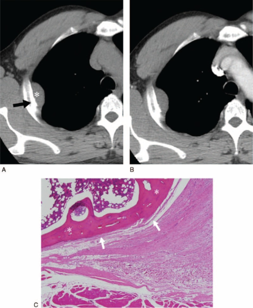
A 52-year-old man with desmoid-type fibromatosis in the chest wall without recurrence (Group 2). This was misdiagnosed as a neurogenic tumor based on preoperative CT but as fibromatosis based on preoperative MR. Axial pre- (A) and contrast-enhanced (B) CT axial images (5-mm reconstruction) show a circumscribed, well-defined mass (∗) without enhancement in the right chest wall. Note the osteoblastic change of the rib (arrow). FDG PET reveals a hypometabolic mass, with a maximal standardized uptake value of 2.3 (not shown). (C) A mass shows abutting the rib (∗) with no evidence of invasion of the cortex of bone and periosteum (arrows, ×40 magnification). CT = computed tomography, MR = magnetic resonance, FDG PET = 18F-fluorodeoxy glucose positron emission tomography.Source: Xu H. et al, Department of Radiology and Research Institute of Radiology, University of Ulsan College of Medicine, Asan Medical Center, Seoul, Korea (HX, HJK, SL, JWL, HNL, MYK); Department of Radiology, The First Affiliated Hospital of Nanjing Medical University, Nanjing, Jiangsu Province, China (HX); Department of Thoracic and Cardiovascular Surgery (DKK); and Pathology, University of Ulsan College of Medicine, Asan Medical Center, Seoul, Korea (JSS).
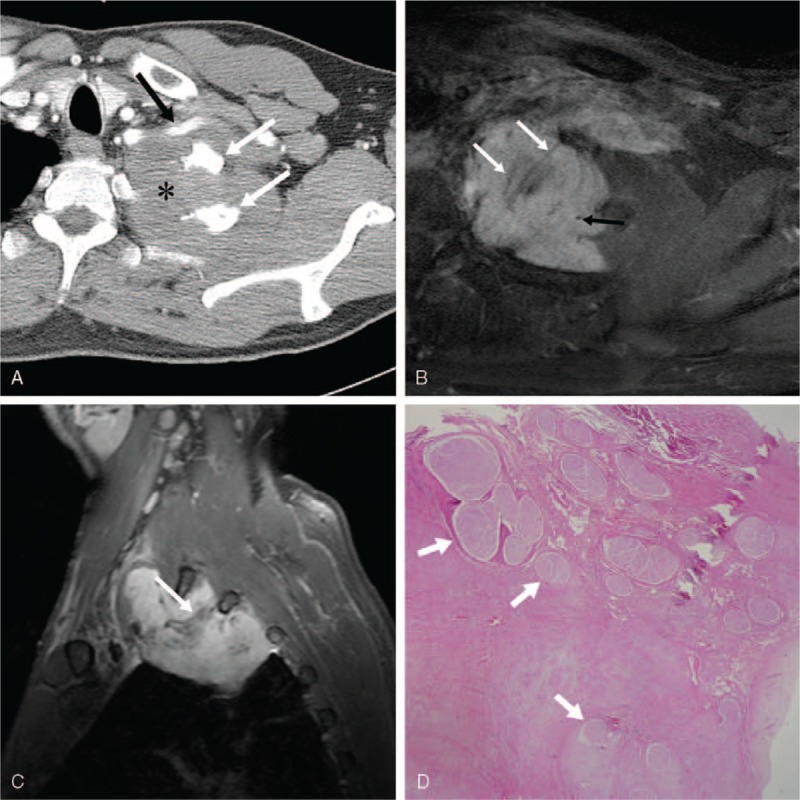
A 47-year-old man presented with desmoid-type fibromatosis in the left supraclavicular area with recurrence (Group 1). It was misdiagnosed as a pancoast tumor due to lung cancer and a sarcoma of mesenchymal origin, based on preoperative CT and MR, respectively. (A) The contrast-enhanced axial (A) CT image (5-mm reconstruction) shows a huge, lobulated, partially ill-defined mass in the supraclavicular area (∗). Note the osteoblastic change of the first and second ribs (white arrows) and suspicion of involvement of the left subclavian artery (black arrow). Note the strong and heterogeneous enhancement with central areas of nonenhancing low signal bands (white arrows) and internal signal voids (black arrow) on enhanced T1-weighted axial (B) and sagittal (C) images. FDG PET revealed a hypermetabolic mass with a maximal standardized uptake value of 4.1 (not shown). (D) The mass was excised. Pathologically, the mass has infiltrated the brachial plexus, and tumor cells surround the nerve bundles (arrows, ×12.5 magnification). CT = computed tomography, MR = magnetic resonance, FDG PET = 18F-fluorodeoxy glucose positron emission tomography.Source: Xu H. et al, Department of Radiology and Research Institute of Radiology, University of Ulsan College of Medicine, Asan Medical Center, Seoul, Korea (HX, HJK, SL, JWL, HNL, MYK); Department of Radiology, The First Affiliated Hospital of Nanjing Medical University, Nanjing, Jiangsu Province, China (HX); Department of Thoracic and Cardiovascular Surgery (DKK); and Pathology, University of Ulsan College of Medicine, Asan Medical Center, Seoul, Korea (JSS).
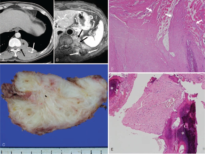
A 43-year-old man with desmoid-type fibromatosis in the mediastinum with recurrence (Group 1). This was misdiagnosed as a neurogenic tumor and as fibrosarcoma on preoperative CT and MR, respectively. (A) The contrast-enhanced axial CT image (5-mm reconstruction) shows a lobulated, well-defined mass (∗) with mild homogeneous enhancement in the left posterior mediastinum. The difference in Hounsfield units between pre- and postenhancement is 7. Note the osteoblastic change of the rib (white arrow) and the neural foraminal widening (black arrow). (B) The tumor shows strong heterogeneous enhancement on an enhanced T1-weighted image. Note the internal nonenhancing low signal bands (white arrows) and internal signal voids (black arrows). (C) Grossly, an ill-defined irregularly-shaped firm mass is present. The cut surface of the mass glistens, and is whitish gray, coarsely trabeculated and partly myxoid, without hemorrhage and necrosis. (D) Tumor cells infiltrate the chest wall muscle (white arrows, ×40 magnification) and (E) also the marrow space of the rib (∗, ×100 magnification). CT = computed tomography, MR = magnetic resonance.Source: Xu H. et al, Department of Radiology and Research Institute of Radiology, University of Ulsan College of Medicine, Asan Medical Center, Seoul, Korea (HX, HJK, SL, JWL, HNL, MYK); Department of Radiology, The First Affiliated Hospital of Nanjing Medical University, Nanjing, Jiangsu Province, China (HX); Department of Thoracic and Cardiovascular Surgery (DKK); and Pathology, University of Ulsan College of Medicine, Asan Medical Center, Seoul, Korea (JSS).
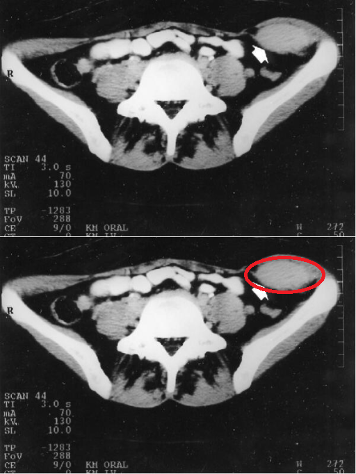
CT Scan with contrast enhancement demonstrates the desmoid tumor originating from the abdominal transversal and internal oblique muscle fascia with inhomogeneous formation (arrow bar).Source: Overhaus M. et al, Department of General-, Visceral-, Thoracic- and Vascular Surgery, Sigmund Freud Str. 25, 53105 Bonn, University of Bonn, Germany
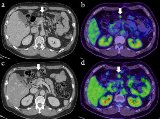
Abdominal computed tomography (CT) and positron emission tomography (PET) of the desmoid tumor. (a) CT shows a solitary and localized tumor in the mesentery of the transverse colon 27 months after gastrectomy (arrow). (b) PET shows radiotracer fluorodeoxyglucose uptake of the tumor (arrow). (c) CT and (d) PET of the tumor after five courses of chemotherapy, showing that no significant clinical response was seen (arrow).Source: Okamura A. et al, Department of Surgery, School of Medicine, Keio University, 35 Shinanomachi, Shinjuku-ku, Tokyo 160-8582, Japan
