Commotio cordis pathophysiology
|
Commotio cordis Microchapters |
|
Diagnosis |
|---|
|
Treatment |
|
Case Studies |
|
Commotio cordis pathophysiology On the Web |
|
American Roentgen Ray Society Images of Commotio cordis pathophysiology |
|
Risk calculators and risk factors for Commotio cordis pathophysiology |
Editor-In-Chief: C. Michael Gibson, M.S., M.D. [1]
Pathophysiology
The following factors increase the chance of commotio cordis[1][2]:
- Direction of impact over the precordium (precise area, angle of impact)
- Total applied energy (area of impact versus energy, i.e., speed of the projectile multiplied by its mass)
- Impact occurring within a specific 10-30 millisecond portion of the cardiac cycle. This period occurs in the ascending phase of the T wave, when the ventricular myocardium is repolarizing, moving from systole to diastole (relaxation).
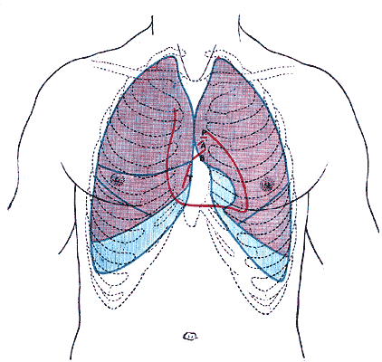
Human adult thorax, showing the outline of the heart (in red). The sensitive zone for mechanical induction of heart rhythm disturbances lies between the 2nd and the 4th ribs, to the left of the sternum

The small window of vulnerability explains why it is a rare event. Considering that the total cardiac cycle has a duration of 1000 milliseconds (for a base cardiac frequency of 60 beats per minute), the probability of a mechanical trauma within the window of vulnerability is 1 to 3% only. That explains also why the heart becomes more vulnerable when it is physically strained by sports activities:
- The increase in heart frequency (exercise tachycardia) may double the probability above (e.g., with 120 beats per minute the cardiac cycle shortens to 500 milliseconds without fundamentally altering the windows of vulnerability size)
- Relative exercise-induced hypoxia and acceleration of the excito- conductive system of the heart make it more susceptible to stretch-induced ventricular fibrillation.
The cellular mechanisms of commotio cordis are still poorly understood, but probably related to the activation of mechano- sensitive proteins, ion channels.
It is estimated that impact energies of at least 50 joules are required to cause cardiac arrest, when applied in the right time and spot of the precordium of an adult. Impacts of up to 130 joules have already been measured with hockey pucks and lacrosse balls, 450 joules in karate punches and an incredible 1028 joules in boxer Rocky Marciano's punch. The 50 joules threshold, however, can be considerably lowered when the victim's heart is under ischemic conditions, such as in coronary artery insufficiency[3].
There is also an upper limit of impact energy applied to the heart; too much energy will create structural damage to the heart muscle as well as causing electrical upset. This condition is referred to as contusio cordis (from Latin for bruising of the heart). On isolated guinea pig hearts, as little as 5 mJ was needed to induce release of creatine kinase, a marker for muscle cell damage.[4] Obviously one should take into account that this figure does not include the dissipation of energy through the chest wall, and is not scaled up for humans, but it is indicative that relatively small amounts of energy are required to reach the heart before physical damage is done.
References
- ↑ Geddes LA, Roeder RA. Evolution of our knowledge of sudden death due to commotio cordis. Am J Emerg Med. 2005 Jan;23(1):67-75. Review. PMID 15672341
- ↑ Kohl P, Nesbitt AD, Cooper PJ, Lei M. Sudden cardiac death by Commotio cordis: role of mechano-electric feedback. Cardiovasc Res. 2001 May;50(2):280-9. Review. PMID 11334832
- ↑ Kohl P, Sachs F & Franz M (eds): Cardiac Mechano-Electric Feedback and Arrhythmias: from Pipette to Patient. Elsevier (Saunders), Philadelphia 2005 ISBN 9781416000341
- ↑ Cooper PJ, Epstein A, Macleod IA, Schaaf ST, Sheldon J, Boulin C, Kohl P. Soft tissue impact characterisation kit (STICK) for ex situ investigation of heart rhythm responses to acute mechanical stimulation. Prog Biophys Mol Biol. 2006 Jan-Apr;90(1-3):444-68. Review. PMID 16125216