Chondrosarcoma CT: Difference between revisions
Jump to navigation
Jump to search
| Line 8: | Line 8: | ||
==Computerized Tomography== | ==Computerized Tomography== | ||
The features seen on CT are the same as on plain film, but are simply better seen. | The features seen on CT are the same as on plain film, but are simply better seen. | ||
*94% of cases demonstrate matrix calcification. | *94% of cases demonstrate [[Calcification|matrix calcification]]. | ||
*Endosteal calcification. | *Endosteal calcification. | ||
*Cortical breach, seen in 88% of longbone chondrosarcoma. | *Cortical breach, seen in 88% of longbone chondrosarcoma. | ||
| Line 21: | Line 21: | ||
Image:Chondrosarcoma 002.jpg|CT images demonstrate a large pelvic chondrosarcoma | Image:Chondrosarcoma 002.jpg|CT images demonstrate a large pelvic chondrosarcoma | ||
</gallery> | </gallery> | ||
==References== | ==References== | ||
Revision as of 17:35, 31 August 2015
|
Chondrosarcoma Microchapters |
|
Diagnosis |
|---|
|
Treatment |
|
Case Studies |
|
Chondrosarcoma CT On the Web |
|
American Roentgen Ray Society Images of Chondrosarcoma CT |
Editor-In-Chief: C. Michael Gibson, M.S., M.D. [1]
Overview
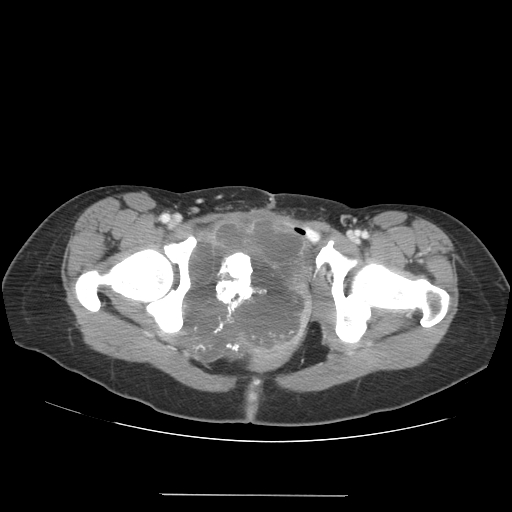
On CT scan chondrosarcoma is characterized by matrix calcification, endosteal calcification, cortical breach, heterogenous contrast enhancement.
Computerized Tomography
The features seen on CT are the same as on plain film, but are simply better seen.
- 94% of cases demonstrate matrix calcification.
- Endosteal calcification.
- Cortical breach, seen in 88% of longbone chondrosarcoma.
- Soft tissue mass: density increases with increased grade of tumor due to increased cellularity.
- Heterogenous contrast enhancement.
(Images shown below are courtesy of RadsWiki)
-
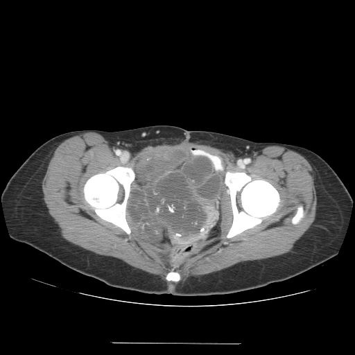
CT images demonstrate a large pelvic chondrosarcoma
-
CT images demonstrate a large pelvic chondrosarcoma

