Cerebral aneurysm pathophysiology
|
Cerebral aneurysm Microchapters |
|
Diagnosis |
|---|
|
Treatment |
|
Case Studies |
|
Cerebral aneurysm pathophysiology On the Web |
|
American Roentgen Ray Society Images of Cerebral aneurysm pathophysiology |
|
Risk calculators and risk factors for Cerebral aneurysm pathophysiology |
Editor-In-Chief: C. Michael Gibson, M.S., M.D. [1] ;Associate Editor(s)-in-Chief: Anika Zahoor M.D.[2]
Overview
Genes involved in the pathogenesis of cerebral aneurysm include [gene1], [gene2], and [gene3]. The pathophysiology of [disease name] depends on the histological subtype.
Pathophysiology
Pathogenesis
Pathogenesis is the mechanism by which a certain factor causes disease (pathos = disease, genesis = development). The term can also be used to describe the development of the disease, whether it is acute, chronic, or recurrent. It can also be used to describe whether the disease causes inflammation, malignancy, necrosis etc.
Template Sentences
IF the pathogenesis of the disease is unclear:
- It is thought that [disease name] is the result of / is mediated by / is produced by / is caused by either hypertension, collagen diseases, or [hypothesis 3].
IF the disease has a known genetic component:
- [Disease name] is transmitted in [mode of genetic transmission] pattern.
- Genes involved in the pathogenesis of [disease name] include [gene1], [gene2], and [gene3].
IF certain pathology findings are characteristic of the disease:
- On gross pathology, [feature1], [feature2], and [feature3] are characteristic findings of [disease name].
- On microscopic histopathological analysis, [feature1], [feature2], and [feature3] are characteristic findings of [disease name].
Other relevant information may include the action of the pathogen on a molecular level, the body’s response, whether or not mutations play a role in the disease development, whether the pathophysiology of the disease is different among subgroups of the disease, etc. Additional template sentences are listed below. Due to the highly variable nature of pathophysiology among various diseases, this list is not comprehensive.
- [Disease or malignancy name] arises from [cell name]s, which are [cell type] cells that are normally involved in [function of cells].
- The development of [disease name] is the result of multiple genetic mutations.
- The progression to [disease name] usually involves the [molecular pathway].
- The pathophysiology of [disease/malignancy] depends on the histological subtype.
- For an example of a pathogenesis section within a pathophysiology page, click here
Pathological Findings
-
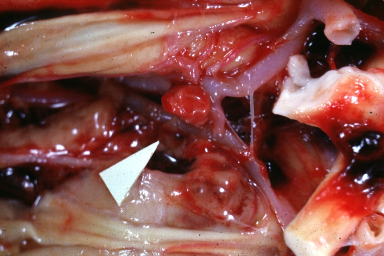
Brain: Berry Aneurysm: Gross, natural color, close-up, an excellent view of typical berry aneurysm located on anterior cerebral artery
-
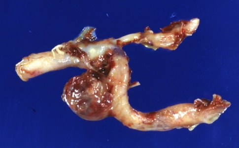
Brain: Berry Aneurysm Ruptured: Gross fixed tissue aneurysm at junction internal carotid and middle cerebral arteries (an excellent close-up view)


References
1.Jung K. H. (2018). New Pathophysiological Considerations on Cerebral Aneurysms. Neurointervention, 13(2), 73–83. https://doi.org/10.5469/neuroint.2018.01011