Brugada syndrome electrocardiographic examples of Type I Brugada syndrome
Editor-In-Chief: C. Michael Gibson, M.S., M.D. [1]
Return to Brugada Syndrome
Overview
Shown below are additional examples of Type I Brugada syndrome.
Examples
- Shown below is an EKG example of Type I Brugada syndrome showing J point elevation, and a right bundle branch block morphology.
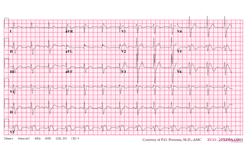
- Shown below is an EKG example of Type I Brugada syndrome showing J point elevation, and a right bundle branch block morphology.
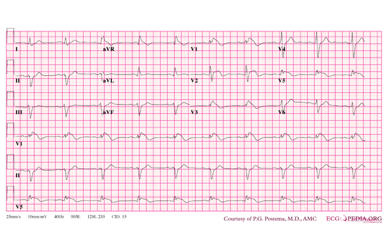
- Shown below is an EKG example of Type I Brugada syndrome showing J point elevation, and a right bundle branch block morphology.
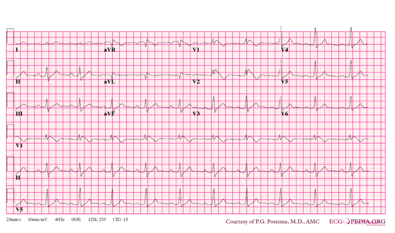
- Shown below is an EKG example of Type I Brugada syndrome showing J point elevation, and a right bundle branch block morphology.
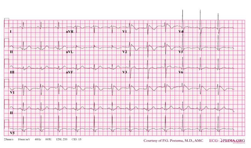
- Shown below is an EKG example of Type I Brugada syndrome showing J point elevation, and a right bundle branch block morphology.
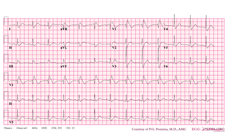
- Shown below is an EKG example of Type I Brugada syndrome showing J point elevation, and a right bundle branch block morphology.
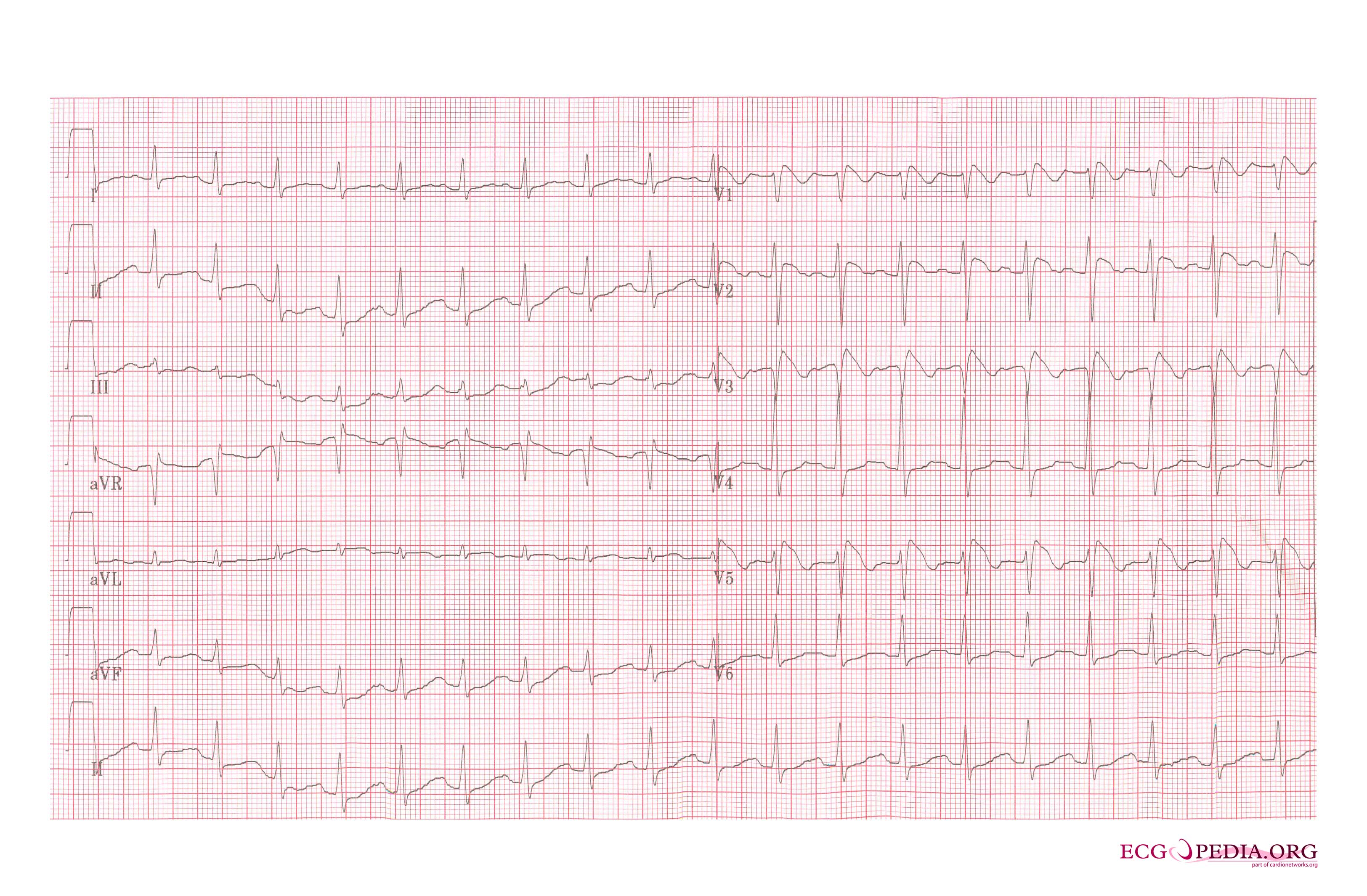
- Shown below is an EKG example of Type I Brugada syndrome showing J point elevation, and a right bundle branch block morphology.
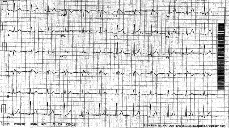
- Shown below is an EKG example of Type I Brugada syndrome showing J point elevation, and a right bundle branch block morphology.
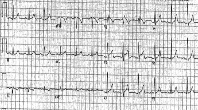
Sources
Copyleft images obtained courtesy of ECGpedia, http://en.ecgpedia.org/index.php?title=Special:NewFiles&dir=prev&offset=20080806182927&limit=500