Brugada syndrome electrocardiographic examples of Type I Brugada syndrome: Difference between revisions
Jump to navigation
Jump to search
| Line 8: | Line 8: | ||
==EKG Examples== | ==EKG Examples== | ||
*Shown below is an EKG example of Type I Brugada syndrome showing J point elevation, and a right bundle branch block morphology. | *Shown below is an EKG example of Type I Brugada syndrome showing [[J point elevation]], and a [[right bundle branch block]] morphology. | ||
[[Image:Brugada_syndrome_type1_example1.png|center|800px]] | [[Image:Brugada_syndrome_type1_example1.png|center|800px]] | ||
---- | ---- | ||
Revision as of 16:33, 24 October 2012
Editor-In-Chief: C. Michael Gibson, M.S., M.D. [1]
For the main page on Brugada Syndrome click here
Overview
Shown below are additional examples of Type I Brugada syndrome.
EKG Examples
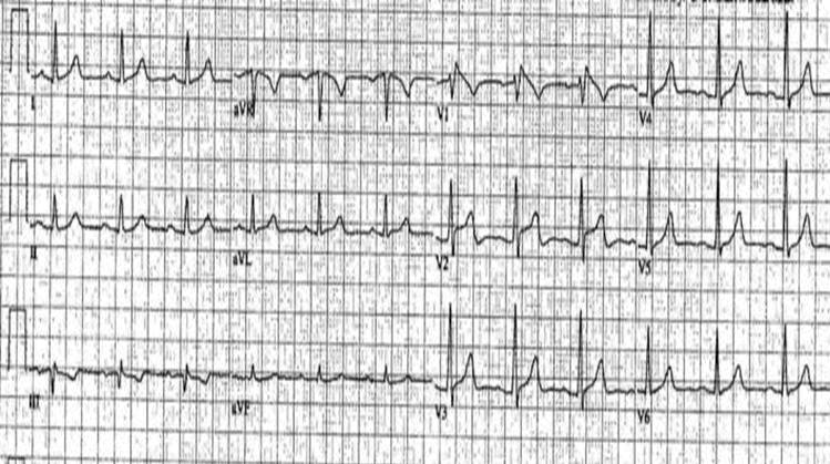
- Shown below is an EKG example of Type I Brugada syndrome showing J point elevation, and a right bundle branch block morphology.
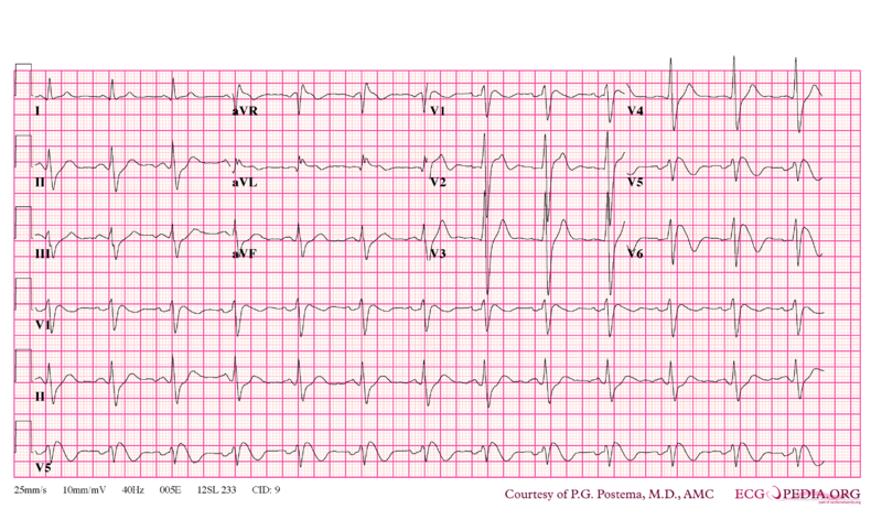
- Shown below is an EKG example of Type I Brugada syndrome showing J point elevation, and a right bundle branch block morphology.
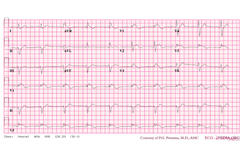
- Shown below is an EKG example of Type I Brugada syndrome showing J point elevation, and a right bundle branch block morphology.
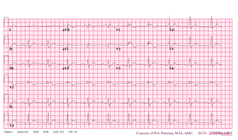
- Shown below is an EKG example of Type I Brugada syndrome showing J point elevation, and a right bundle branch block morphology.
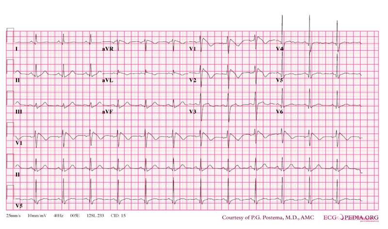
- Shown below is an EKG example of Type I Brugada syndrome showing J point elevation, and a right bundle branch block morphology.
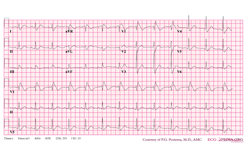
- Shown below is an EKG example of Type I Brugada syndrome showing J point elevation, and a right bundle branch block morphology. It was obtained during ajmaline testing.
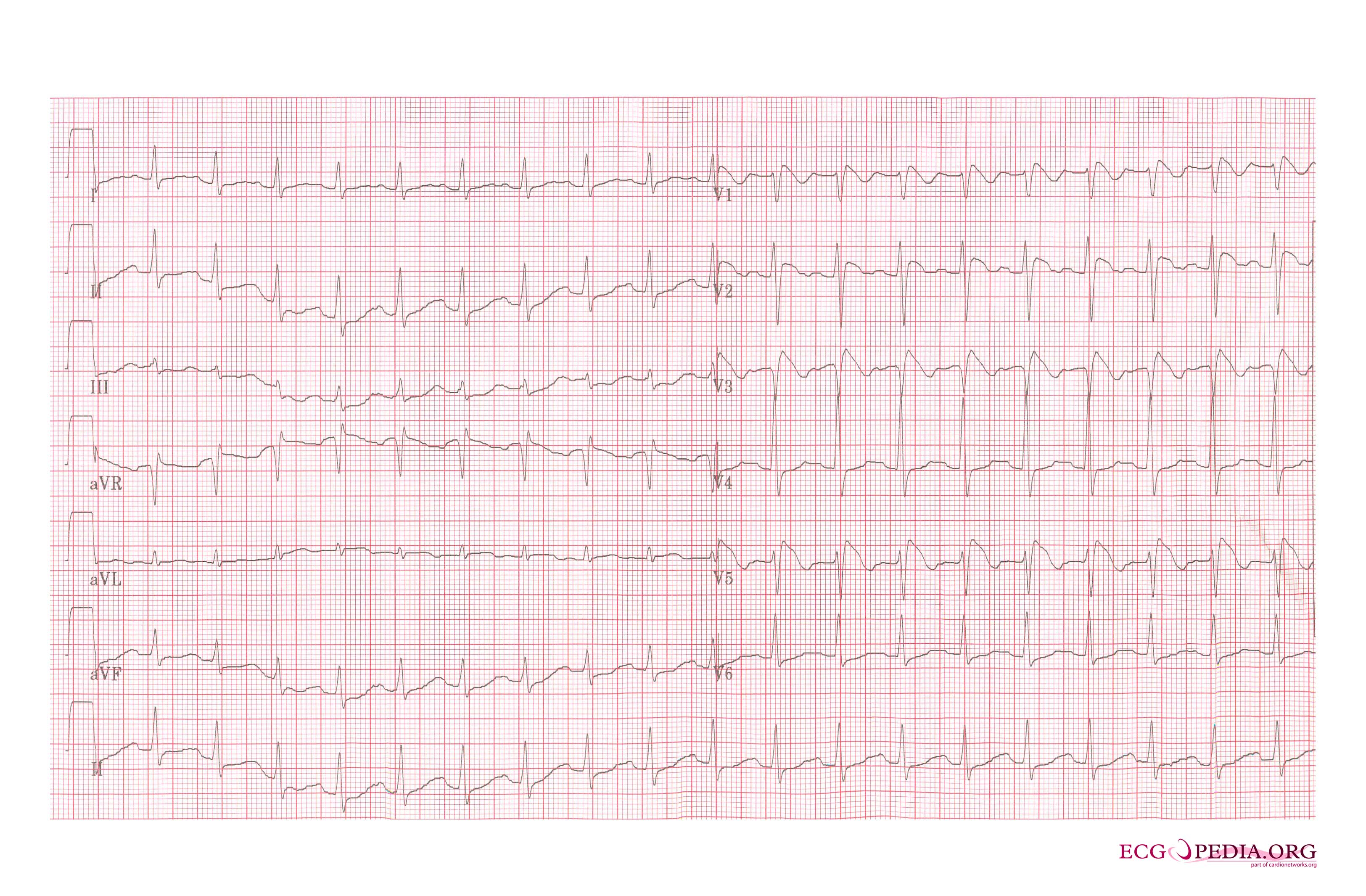
- Shown below is an EKG example of Type I Brugada syndrome showing J point elevation, and a right bundle branch block morphology.
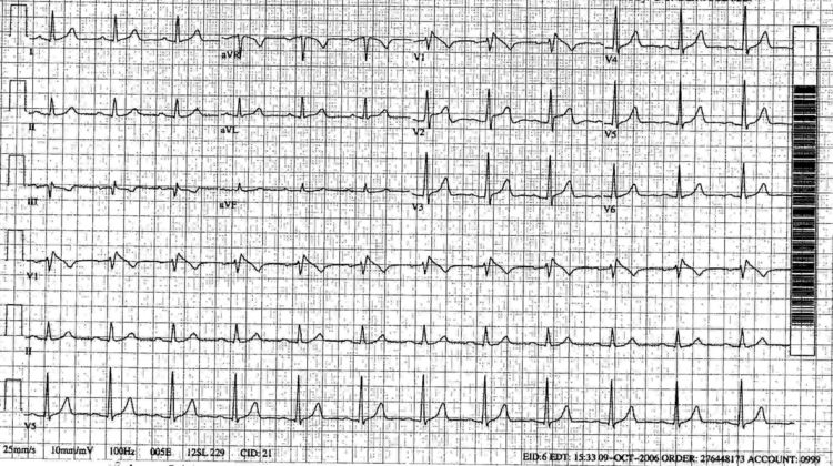
- Shown below is an EKG example of Type I Brugada syndrome showing J point elevation, and a right bundle branch block morphology.
Sources
Copyleft images obtained courtesy of ECGpedia, http://en.ecgpedia.org/index.php?title=Special:NewFiles&offset=&limit=500