* Pneumonia was first recognized by Hippocrates. It was first identified and described by Laennec in 1819.
* Pneumonia was first recognized by Hippocrates. It was first identified and described by Laennec in 1819.
* In 1842, Rokitansky differentiated Pneumonia into Bronchopneumonia and Lobar Pneumonia.
* In 1842, Rokitansky differentiated Pneumonia into Bronchopneumonia and Lobar Pneumonia.
== Classification[edit | edit source] ==
== Classification ==
* Pneumonia may be classified according to anatomic distribution of consolidation into two subtypes/groups:
* Pneumonia may be classified according to anatomic distribution of consolidation into two subtypes/groups:
** Lobar
** Lobar
** Lobular (Bronchopneumonia)
** Lobular (Bronchopneumonia)
== Pathophysiology[edit | edit source] ==
== Pathophysiology ==
* The pathogenesis of Bronchopneumonia is characterized by inflammation of lung parenchyma.
* The pathogenesis of Bronchopneumonia is characterized by inflammation of lung parenchyma.
* On gross pathology, multiple foci of consolidation is a characteristic feature of Bronchopneumonia. They are present bilaterally, most commonly in the basal lobes. These lesions are 2-4 cm in diameter, grey-yellow, dry, often centered by a [[bronchia]], are poorly delimited and have the tendency to confluence, especially in children.
* On gross pathology, multiple foci of consolidation is a characteristic feature of Bronchopneumonia. They are present bilaterally, most commonly in the basal lobes. These lesions are 2-4 cm in diameter, grey-yellow, dry, often centered by a [[bronchia]], are poorly delimited and have the tendency to confluence, especially in children.
Line 15:
Line 15:
* Bronchopneumonia is usually associated with infections due to gram-negative bacteria, ''Staphylococcus aureus'' and some fungi.
* Bronchopneumonia is usually associated with infections due to gram-negative bacteria, ''Staphylococcus aureus'' and some fungi.
== Clinical Features[edit | edit source] ==
== Clinical Features ==
* Common clinical findings in Bronchopneumonia include cough, fever, chills, dyspnea, pleuritic chest pain and sputum production. However, many of these features may be absent in older patients.
* Common clinical findings in Bronchopneumonia include cough, fever, chills, dyspnea, pleuritic chest pain and sputum production. However, many of these features may be absent in older patients.
* Bronchopneumonia can also case Gastrointestinal symptoms such as nausea, vomiting and diarrhea.
* Bronchopneumonia can also case Gastrointestinal symptoms such as nausea, vomiting and diarrhea.
* Older patients may also present with altered mental status.
* Older patients may also present with altered mental status.
== Differentiating [disease name] from other Diseases[edit | edit source] ==
== Differentiating Bronchopneumonia from other Diseases ==
* Lobular pneumonia must be differentiated from other diseases that cause similar clinical symptoms and interstitial infiltrates on chest x-ray such as:
* Lobular pneumonia must be differentiated from other diseases that cause similar clinical symptoms and interstitial infiltrates on chest x-ray such as:
:* Lobar pneumonia
:* Lobar pneumonia
Line 26:
Line 26:
:* Other types of pneumonias such as: cryptogenic pneumonia.
:* Other types of pneumonias such as: cryptogenic pneumonia.
== Epidemiology and Demographics[edit | edit source] ==
== Epidemiology and Demographics ==
* The rate of Community acquired pneumonia is approximately 5.16-7.06 cases per 1000 individuals per year.
* The rate of Community acquired pneumonia is approximately 5.16-7.06 cases per 1000 individuals per year.
Line 40:
Line 40:
* Bronchopneumonia is more commonly observed in Black persons than caucasians.
* Bronchopneumonia is more commonly observed in Black persons than caucasians.
== Risk Factors[edit | edit source] ==
== Risk Factors ==
* Common risk factors in the development of Bronchopneumonia are Influenza infection, Alcohol abuse, Hyposplenism/splenectomy, smoking, COPD/Asthma and Immunocompromise. Additional risk factors include, homelessness, incarceration, pregnancy, crack cocaine use, opioid use and occupational welding.
* Common risk factors in the development of Bronchopneumonia are Influenza infection, Alcohol abuse, Hyposplenism/splenectomy, smoking, COPD/Asthma and Immunocompromise. Additional risk factors include, homelessness, incarceration, pregnancy, crack cocaine use, opioid use and occupational welding.
* Risk factors for a complicated course include, older age, preexisting lung condition, immunodeficiency/AIDS, and acquisition of a nosocomial infection.
* Risk factors for a complicated course include, older age, preexisting lung condition, immunodeficiency/AIDS, and acquisition of a nosocomial infection.
== Natural History, Complications and Prognosis[edit | edit source] ==
== Natural History, Complications and Prognosis ==
* Early clinical features include sudden fever, chills, cough and chest pain.<ref name="Mackenzie20165" />
* Early clinical features include sudden fever, chills, cough and chest pain.<ref name="Mackenzie20165" />
* If left untreated, patients with Bronchopneumonia may progress to develop tachypnea and increasing systemic toxicity. They may also progress to develop Lobar pneumonia.
* If left untreated, patients with Bronchopneumonia may progress to develop tachypnea and increasing systemic toxicity. They may also progress to develop Lobar pneumonia.
* Common complications of Bronchopneumonia include parapneumonic effusion, empyema, necrotizing pneumonia, lung abscess and metastatic infections such as endocarditis, septic arthritis, peritonitis, pericarditis and meningitis, and other cardiac complications.
* Common complications of Bronchopneumonia include parapneumonic effusion, empyema, necrotizing pneumonia, lung abscess and metastatic infections such as endocarditis, septic arthritis, peritonitis, pericarditis and meningitis, and other cardiac complications.
== Diagnosis[edit | edit source] ==
== Diagnosis ==
=== Diagnostic Criteria[edit | edit source] ===
=== Diagnostic Study of choice ===
* The diagnosis is made when clinical and radiological evidence suggests the presence of Bronchopneumonia.
* The diagnosis is made when clinical and radiological evidence suggests the presence of Bronchopneumonia.
=== Symptoms[edit | edit source] ===
=== Symptoms ===
*Symptoms of Bronchopneumonia may include the following:
*Symptoms of Bronchopneumonia may include the following:
** Fever
** Fever
Line 60:
Line 60:
** Chest Pain
** Chest Pain
** Shortness of breath
** Shortness of breath
=== Physical Examination[edit | edit source] ===
=== Physical Examination ===
* Physical examination may be remarkable for:
* Physical examination may be remarkable for:
** Fever
** Fever
Line 71:
Line 71:
*** Tactile fremitus
*** Tactile fremitus
*** Egophony
*** Egophony
=== Laboratory Findings[edit | edit source] ===
=== Laboratory Findings ===
* There are no specific laboratory findings associated with Bronchopneumonia.
* There are no specific laboratory findings associated with Bronchopneumonia.
Line 77:
Line 77:
* An elevated concentration of ESR or CRP is a non-specific indication of inflammation in the body.
* An elevated concentration of ESR or CRP is a non-specific indication of inflammation in the body.
=== Imaging Findings[edit | edit source] ===
=== Imaging Findings ===
* Chest x-ray is the imaging modality of choice for Bronchopneumonia.<ref name="pmid17278083">{{cite journal |vauthors=Mandell LA, Wunderink RG, Anzueto A, Bartlett JG, Campbell GD, Dean NC, Dowell SF, File TM, Musher DM, Niederman MS, Torres A, Whitney CG |title=Infectious Diseases Society of America/American Thoracic Society consensus guidelines on the management of community-acquired pneumonia in adults |journal=Clin. Infect. Dis. |volume=44 Suppl 2 |issue= |pages=S27–72 |date=March 2007 |pmid=17278083 |doi=10.1086/511159 |url=}}</ref>
* Chest x-ray is the imaging modality of choice for Bronchopneumonia.
* On chest x-ray, Bronchopneumonia is characterized by peribronchial thickening and poorly defined air-space opacities. Inhomogeneous patchy areas of consolidation involving several lobes reflect more severe disease. When confluent, bronchopneumonia may resemble lobar pneumonia.<ref name="pmid172780832">{{cite journal |vauthors=Mandell LA, Wunderink RG, Anzueto A, Bartlett JG, Campbell GD, Dean NC, Dowell SF, File TM, Musher DM, Niederman MS, Torres A, Whitney CG |title=Infectious Diseases Society of America/American Thoracic Society consensus guidelines on the management of community-acquired pneumonia in adults |journal=Clin. Infect. Dis. |volume=44 Suppl 2 |issue= |pages=S27–72 |date=March 2007 |pmid=17278083 |doi=10.1086/511159 |url=}}</ref>
* On chest x-ray, Bronchopneumonia is characterized by peribronchial thickening and poorly defined air-space opacities. Inhomogeneous patchy areas of consolidation involving several lobes reflect more severe disease. When confluent, bronchopneumonia may resemble lobar pneumonia.
* In the case of a negative chest x-ray and a high clinical suspicion, HRCT scan may be used to confirm the diagnosis as it has a higher sensitivity and accuracy in detecting lesions and anatomical changes.
* In the case of a negative chest x-ray and a high clinical suspicion, HRCT scan may be used to confirm the diagnosis as it has a higher sensitivity and accuracy in detecting lesions and anatomical changes.
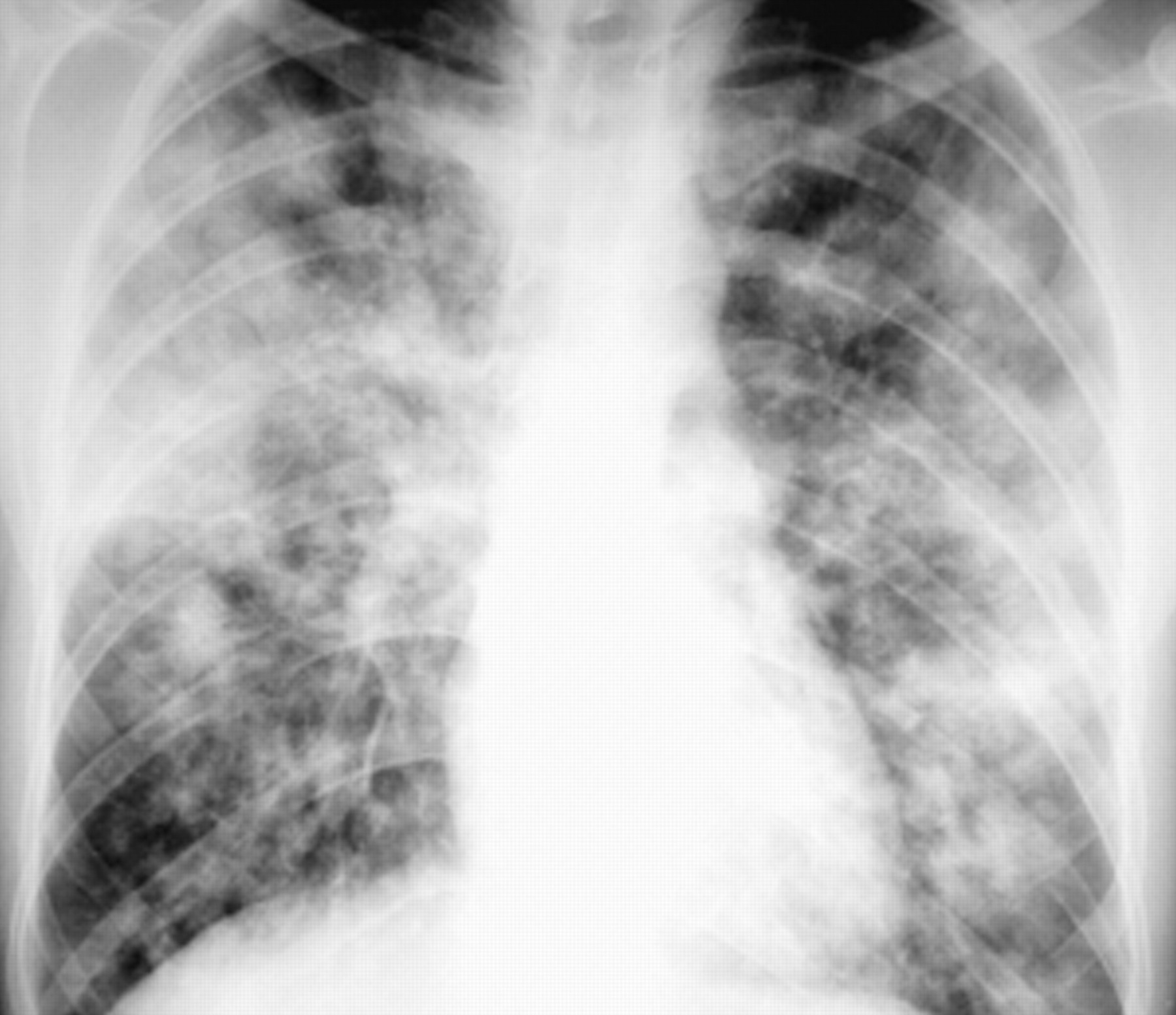
*[[File:Bronchopneumonia caused by aspergillus.jpg|thumb|Posteroanterior chest radiograph reveals bilateral nonsegmental consolidations in the lingula and in the right upper and lower lobes. ''Aspergillus fumigatus'' was recovered from the sputum. ]]In case of emergency where chest x-ray and HRCT cannot be performed, lung ultrasound performed by an experienced physician can yield findings.
*[[File:Bronchopneumonia caused by aspergillus.jpg|thumb|Posteroanterior chest radiograph reveals bilateral nonsegmental consolidations in the lingula and in the right upper and lower lobes. ''Aspergillus fumigatus'' was recovered from the sputum. ]]In case of emergency where chest x-ray and HRCT cannot be performed, lung ultrasound performed by an experienced physician can yield findings.
=== Other Diagnostic Studies[edit | edit source] ===
=== Other Diagnostic Studies ===
* Microbial analysis in Bronchopneumonia can be performed using techniques such as Blood culture, Sputum analysis, PCR and Urine antigen detection, however, pathogens are not commonly identified and empiric treatment should be started once the diagnosis is made.<ref name="pmid172780833">{{cite journal |vauthors=Mandell LA, Wunderink RG, Anzueto A, Bartlett JG, Campbell GD, Dean NC, Dowell SF, File TM, Musher DM, Niederman MS, Torres A, Whitney CG |title=Infectious Diseases Society of America/American Thoracic Society consensus guidelines on the management of community-acquired pneumonia in adults |journal=Clin. Infect. Dis. |volume=44 Suppl 2 |issue= |pages=S27–72 |date=March 2007 |pmid=17278083 |doi=10.1086/511159 |url=}}</ref>
* Microbial analysis in Bronchopneumonia can be performed using techniques such as Blood culture, Sputum analysis, PCR and Urine antigen detection, however, pathogens are not commonly identified and empiric treatment should be started once the diagnosis is made.
== Treatment[edit | edit source] ==
== Treatment ==
=== Medical Therapy[edit | edit source] ===
=== Medical Therapy ===
* The mainstay of therapy for Bronchopneumonia is antibiotics and supportive care.
* The mainstay of therapy for Bronchopneumonia is antibiotics and supportive care.
* Choice of antibiotics is dependent on epidemiology of microbes, resistance and patients' co-morbidities and severity of illness.
* Choice of antibiotics is dependent on epidemiology of microbes, resistance and patients' co-morbidities and severity of illness.
Line 94:
Line 94:
* Response to antibiotics can be monitored with clinical improvement, serum inflammatory markers and chest x-ray findings. However, most non-complicated pneumonias are treated out-patient and only require two follow up treatments to clinically determine improvement and resolution of the pneumonia, respectively. Follow-up chest x-rays are only required in male patients over the age of 50 years and smokers.
* Response to antibiotics can be monitored with clinical improvement, serum inflammatory markers and chest x-ray findings. However, most non-complicated pneumonias are treated out-patient and only require two follow up treatments to clinically determine improvement and resolution of the pneumonia, respectively. Follow-up chest x-rays are only required in male patients over the age of 50 years and smokers.
=== Prevention[edit | edit source] ===
=== Prevention ===
* Effective measures for the primary prevention of Bronchopneumonia include vaccination against influenza and pneumococcal antigens, and smoking cessation. Pneumococcal vaccination is indicated in patients over the age of 65 years.
* Effective measures for the primary prevention of Bronchopneumonia include vaccination against influenza and pneumococcal antigens, and smoking cessation. Pneumococcal vaccination is indicated in patients over the age of 65 years.
Pneumonia was first recognized by Hippocrates. It was first identified and described by Laennec in 1819.
In 1842, Rokitansky differentiated Pneumonia into Bronchopneumonia and Lobar Pneumonia.
Classification
Pneumonia may be classified according to anatomic distribution of consolidation into two subtypes/groups:
Lobar
Lobular (Bronchopneumonia)
Pathophysiology
The pathogenesis of Bronchopneumonia is characterized by inflammation of lung parenchyma.
On gross pathology, multiple foci of consolidation is a characteristic feature of Bronchopneumonia. They are present bilaterally, most commonly in the basal lobes. These lesions are 2-4 cm in diameter, grey-yellow, dry, often centered by a bronchia, are poorly delimited and have the tendency to confluence, especially in children.
On microscopic histopathological analysis, a focus of inflammatory condensation, centered by a bronchiola with acute bronchiolitis is a characteristic finding in Bronchopneumonia. In addition, alveolar lumens surrounding the bronchia are filled with neutrophils and suppurative exudate("leukocytic alveolitis"), massive congestion is present and inflammatory foci are separated by normal, aerated parenchyma.
Bronchopneumonia is most commonly caused by pneumococcal serotypes 3, 7,8,10,18 and 20.
Common mechanisms in development of pneumonia include, micro-aspiration, hematogenous spread, spread from a contiguous focus and macro-aspiration.
Bronchopneumonia is usually associated with infections due to gram-negative bacteria, Staphylococcus aureus and some fungi.
Clinical Features
Common clinical findings in Bronchopneumonia include cough, fever, chills, dyspnea, pleuritic chest pain and sputum production. However, many of these features may be absent in older patients.
Bronchopneumonia can also case Gastrointestinal symptoms such as nausea, vomiting and diarrhea.
Older patients may also present with altered mental status.
Differentiating Bronchopneumonia from other Diseases
Lobular pneumonia must be differentiated from other diseases that cause similar clinical symptoms and interstitial infiltrates on chest x-ray such as:
Lobar pneumonia
Non-infectious lung conditions such as: Hypersensitivity pneumonitis, Collagen vascular disease, Asbestosis, Drug toxicities, Pulmonary fibrosis, Pulmonary edema, Pulmonary embolism and neoplastic lesions.
Other types of pneumonias such as: cryptogenic pneumonia.
Epidemiology and Demographics
The rate of Community acquired pneumonia is approximately 5.16-7.06 cases per 1000 individuals per year.
Age[edit | edit source]
Patients of all age groups may develop Bronchopneumonia.
Bronchopneumonia is more commonly observed among elderly patients.
Gender[edit | edit source]
Brochopneumonia is more commonly observed in men than women.
Race[edit | edit source]
Bronchopneumonia is more commonly observed in Black persons than caucasians.
Risk Factors
Common risk factors in the development of Bronchopneumonia are Influenza infection, Alcohol abuse, Hyposplenism/splenectomy, smoking, COPD/Asthma and Immunocompromise. Additional risk factors include, homelessness, incarceration, pregnancy, crack cocaine use, opioid use and occupational welding.
Risk factors for a complicated course include, older age, preexisting lung condition, immunodeficiency/AIDS, and acquisition of a nosocomial infection.
Natural History, Complications and Prognosis
Early clinical features include sudden fever, chills, cough and chest pain.[1]
If left untreated, patients with Bronchopneumonia may progress to develop tachypnea and increasing systemic toxicity. They may also progress to develop Lobar pneumonia.
Common complications of Bronchopneumonia include parapneumonic effusion, empyema, necrotizing pneumonia, lung abscess and metastatic infections such as endocarditis, septic arthritis, peritonitis, pericarditis and meningitis, and other cardiac complications.
Diagnosis
Diagnostic Study of choice
The diagnosis is made when clinical and radiological evidence suggests the presence of Bronchopneumonia.
Symptoms
Symptoms of Bronchopneumonia may include the following:
Fever
Chills
Cough
Chest Pain
Shortness of breath
Physical Examination
Physical examination may be remarkable for:
Fever
Respiratory rate >24 breaths/min (Tachypnea)
Tachycardia
Chest Examination:
Audible crackles
Decreased or bronchial breath sounds
Dullness to percussion in areas of consolidation
Tactile fremitus
Egophony
Laboratory Findings
There are no specific laboratory findings associated with Bronchopneumonia.
A Leukocytosis (15000-30000 per mm3) with a left ward shift on a blood test can aid in diagnosis of Bronchopneumonia.
An elevated concentration of ESR or CRP is a non-specific indication of inflammation in the body.
Imaging Findings
Chest x-ray is the imaging modality of choice for Bronchopneumonia.
On chest x-ray, Bronchopneumonia is characterized by peribronchial thickening and poorly defined air-space opacities. Inhomogeneous patchy areas of consolidation involving several lobes reflect more severe disease. When confluent, bronchopneumonia may resemble lobar pneumonia.
In the case of a negative chest x-ray and a high clinical suspicion, HRCT scan may be used to confirm the diagnosis as it has a higher sensitivity and accuracy in detecting lesions and anatomical changes.
Posteroanterior chest radiograph reveals bilateral nonsegmental consolidations in the lingula and in the right upper and lower lobes. Aspergillus fumigatus was recovered from the sputum.In case of emergency where chest x-ray and HRCT cannot be performed, lung ultrasound performed by an experienced physician can yield findings.
Other Diagnostic Studies
Microbial analysis in Bronchopneumonia can be performed using techniques such as Blood culture, Sputum analysis, PCR and Urine antigen detection, however, pathogens are not commonly identified and empiric treatment should be started once the diagnosis is made.
Treatment
Medical Therapy
The mainstay of therapy for Bronchopneumonia is antibiotics and supportive care.
Choice of antibiotics is dependent on epidemiology of microbes, resistance and patients' co-morbidities and severity of illness.
In patients without co-morbidities Macrolides such as Azithromycin and Clarithromycin can be used. In the case of Macrolide resistant pneumonias and patients with multiple co-morbidities, Doxycycline, Amoxicillin-Clavulanate, and Cephalosporins such as Cefpodoxime and Cefuroxime may be used.
Response to antibiotics can be monitored with clinical improvement, serum inflammatory markers and chest x-ray findings. However, most non-complicated pneumonias are treated out-patient and only require two follow up treatments to clinically determine improvement and resolution of the pneumonia, respectively. Follow-up chest x-rays are only required in male patients over the age of 50 years and smokers.
Prevention
Effective measures for the primary prevention of Bronchopneumonia include vaccination against influenza and pneumococcal antigens, and smoking cessation. Pneumococcal vaccination is indicated in patients over the age of 65 years.