Bronchogenic cyst pathophysiology
|
Bronchogenic cyst Microchapters |
|
Diagnosis |
|---|
|
Treatment |
|
Case Studies |
|
Bronchogenic cyst pathophysiology On the Web |
|
American Roentgen Ray Society Images of Bronchogenic cyst pathophysiology |
|
Risk calculators and risk factors for Bronchogenic cyst pathophysiology |
Editor-In-Chief: C. Michael Gibson, M.S., M.D. [1]
Overview
Pathophysiology
Bronchogenic cysts originate from anomalous development of the ventral foregut and can be found throughout the tracheoesophageal distribution including perihilar or intraparenchymal areas. they are usually single but may be multiple.
The most common location to find the anomaly is in the carina. The rarest locations include the interatrial septum, neck, abdomen, and retroperitoneal space.
If the cyst compresses a vital respiratory or cardiac structure, it can be symptomatic and life threatening.
Gross Pathology
Images shown below are courtesy of Professor Peter Anderson DVM PhD and published with permission. © PEIR, University of Alabama at Birmingham, Department of Pathology
-
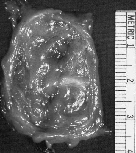
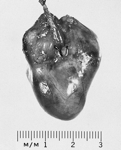
Lower Respiratory Tract: Bronchogenic cysts are grossly nondescript, usually unilocular cavities containing mucus or mucopurulent material with a wall that varies in thickness and may contain cartilage.
-
Lower Respiratory Tract: Bronchogenic cysts are grossly nondescript, usually unilocular cavities containing mucus or mucopurulent material with a wall that varies in thickness and may contain cartilage.


-
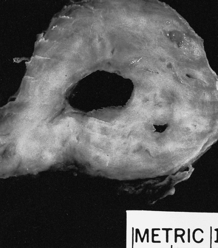
Mediastinum: Bronchogenic cyst; A thin-walled cyst which was present in the posterior mediastinum is distended with slightly cloudy somewhat mucinous fluid.
-
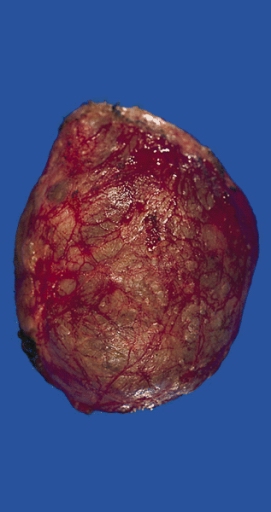
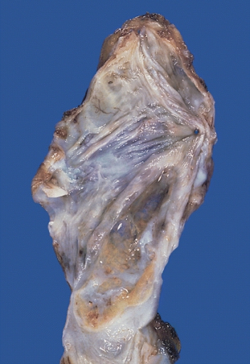
Heart-Great Vessels: Bronchogenic cyst; This example was removed from the epicardial surface of a 13-year-old.


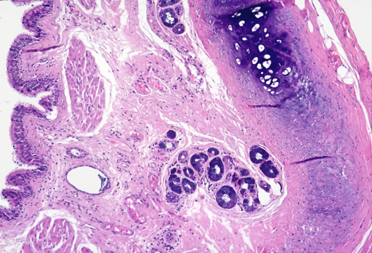
Microscopic Pathology
-
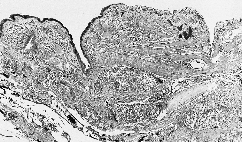
Lower Respiratory Tract: Intrathoracic bronchogenic cyst; Histologically, smooth muscle bundles, minor salivary gland type tissue, and cartilage can be seen in the wall of the cyst.
-
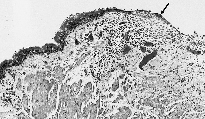
Lower Respiratory Tract: Intrathoracic bronchogenic cyst; The lining is typically pseudostratified ciliated columnar epithelium which may show foci of squamous metaplasia at sites of inflammation (arrow).


-
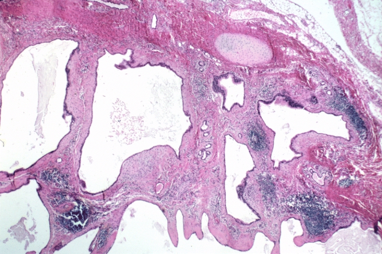
Lower Respiratory Tract: Bronchogenic cyst; There is a cyst lined by bronchiolar epithelium and thick fascicles of smooth muscle in the wall. Islands of cartilage and submucosal salivary glands are lacking and the designation "foregut cyst consistent with bronchogenic cyst" might be more appropriate.
-
Lower Respiratory Tract: Bronchogenic cyst; There is a cyst lined by bronchiolar epithelium and thick fascicles of smooth muscle in the wall. Islands of cartilage and submucosal salivary glands are lacking and the designation "foregut cyst consistent with bronchogenic cyst" might be more appropriate.


-
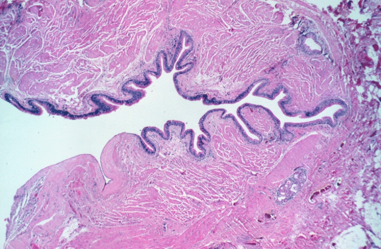
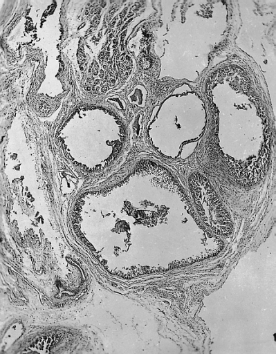
Lower Respiratory Tract: Bronchogenic cyst; There is a multiloculated cyst lined by bronchiolar epithelium. A small island of cartilage is present at the upper right.
-
Heart-Great Vessels: Bronchogenic cyst; The lining cells are cuboidal, and the cysts are surrounded by muscle and may contain underlying seromucous glands.


-
Mediastinum: Bronchogenic cyst; The inner surface is smooth and trabeculated in areas.
-
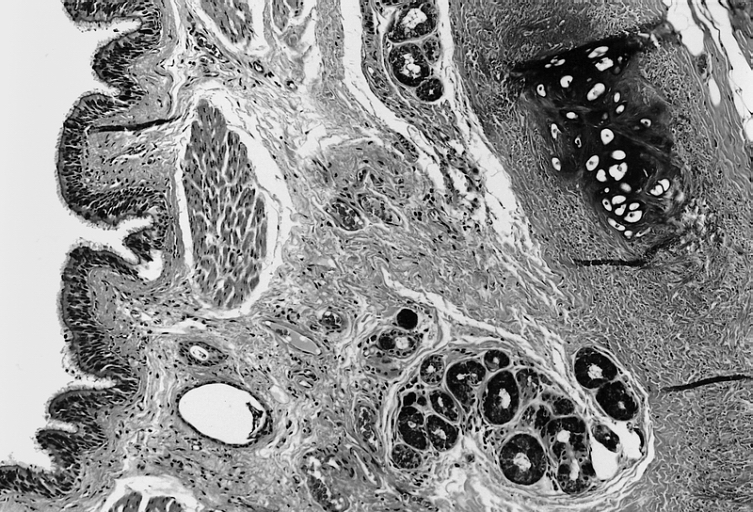
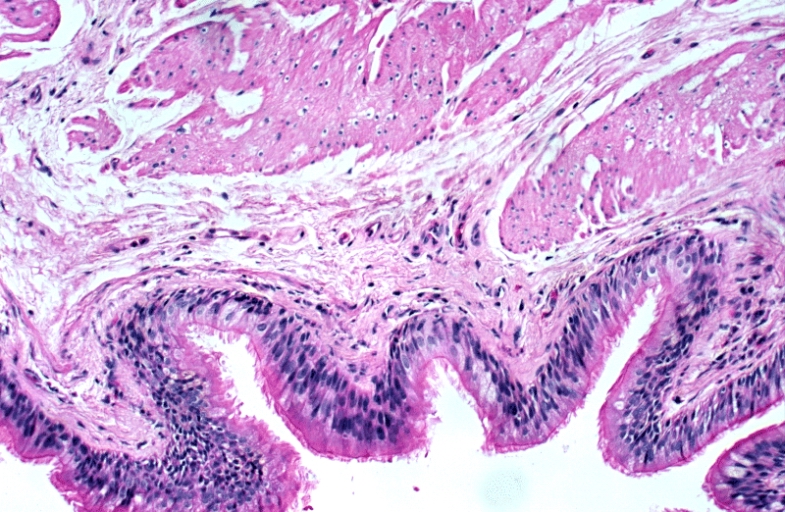
Mediastinum: Bronchogenic cyst; The cyst is lined by pseudostratified ciliated columnar epithelial cells and the wall contains accessory glands, layers of smooth muscle, and cartilage.
-
Mediastinum: Bronchogenic cyst; The cyst is lined by pseudostratified ciliated columnar epithelial cells and the wall contains accessory glands, layers of smooth muscle, and cartilage.


