Blepharitis physical examination: Difference between revisions
No edit summary |
m (Bot: Removing from Primary care) |
||
| (48 intermediate revisions by 6 users not shown) | |||
| Line 1: | Line 1: | ||
__NOTOC__ | __NOTOC__ | ||
{{Blepharitis}} | {{Blepharitis}} | ||
{{CMG}} {{AE}} {{SaraM}} | {{CMG}}; {{AE}} {{SaraM}} | ||
==Overview== | ==Overview== | ||
Physical examination of patients with blepharitis is usually remarkable for irritated eyelid edges, hard crusting of the lashes | Physical examination of patients with blepharitis is usually remarkable for irritated eyelid edges, hard crusting of the lashes, [[entropion]], [[ectropion]], [[poliosis]], and diffuse [[conjunctival injection]].<ref name="pmid3957566">{{cite journal| author=Dougherty JM, McCulley JP| title=Bacterial lipases and chronic blepharitis. | journal=Invest Ophthalmol Vis Sci | year= 1986 | volume= 27 | issue= 4 | pages= 486-91 | pmid=3957566 | doi= | pmc= | url=http://www.ncbi.nlm.nih.gov/entrez/eutils/elink.fcgi?dbfrom=pubmed&tool=sumsearch.org/cite&retmode=ref&cmd=prlinks&id=3957566 }} </ref><ref name="pmid13274422">{{cite journal| author=THYGESON P, VAUGHAN DG| title=Seborrheic blepharitis. | journal=Trans Am Ophthalmol Soc | year= 1954 | volume= 52 | issue= | pages= 173-88 | pmid=13274422 | doi= | pmc=1312591 | url=http://www.ncbi.nlm.nih.gov/entrez/eutils/elink.fcgi?dbfrom=pubmed&tool=sumsearch.org/cite&retmode=ref&cmd=prlinks&id=13274422 }} </ref> | ||
==Physical Examination== | ==Physical Examination== | ||
=== | ===Skin=== | ||
Common skin examination findings of blepharitis include:<ref name="pmid3957566">{{cite journal| author=Dougherty JM, McCulley JP| title=Bacterial lipases and chronic blepharitis. | journal=Invest Ophthalmol Vis Sci | year= 1986 | volume= 27 | issue= 4 | pages= 486-91 | pmid=3957566 | doi= | pmc= | url=http://www.ncbi.nlm.nih.gov/entrez/eutils/elink.fcgi?dbfrom=pubmed&tool=sumsearch.org/cite&retmode=ref&cmd=prlinks&id=3957566 }} </ref><ref name="pmid13274422">{{cite journal| author=THYGESON P, VAUGHAN DG| title=Seborrheic blepharitis. | journal=Trans Am Ophthalmol Soc | year= 1954 | volume= 52 | issue= | pages= 173-88 | pmid=13274422 | doi= | pmc=1312591 | url=http://www.ncbi.nlm.nih.gov/entrez/eutils/elink.fcgi?dbfrom=pubmed&tool=sumsearch.org/cite&retmode=ref&cmd=prlinks&id=13274422 }} </ref><ref name="pmid19383269">{{cite journal| author=Lemp MA, Nichols KK| title=Blepharitis in the United States 2009: a survey-based perspective on prevalence and treatment. | journal=Ocul Surf | year= 2009 | volume= 7 | issue= 2 Suppl | pages= S1-S14 | pmid=19383269 | doi= | pmc= | url=http://www.ncbi.nlm.nih.gov/entrez/eutils/elink.fcgi?dbfrom=pubmed&tool=sumsearch.org/cite&retmode=ref&cmd=prlinks&id=19383269 }} </ref> | |||
Facial flushing | *Scalp or facial skin itching and flaking (suggestive of [[seborrheic dermatitis]]) | ||
*Facial flushing and [[Telangiectasia]] on cheeks and nose (suggestive of [[acne rosacea]]) | |||
=== | |||
===Eye=== | |||
====Gross Eye Examination==== | |||
Common eye examination findings of blepharitis include:<ref name="pmid13274422">{{cite journal| author=THYGESON P, VAUGHAN DG| title=Seborrheic blepharitis. | journal=Trans Am Ophthalmol Soc | year= 1954 | volume= 52 | issue= | pages= 173-88 | pmid=13274422 | doi= | pmc=1312591 | url=http://www.ncbi.nlm.nih.gov/entrez/eutils/elink.fcgi?dbfrom=pubmed&tool=sumsearch.org/cite&retmode=ref&cmd=prlinks&id=13274422 }} </ref><ref name=Blepharitis-1> Blepharitis. American Academy of Ophthalmology/eyewiki (2014). http://eyewiki.org/Blepharitis Accessed on July 14, 2016 </ref><ref name=Blepharitis-3> Blepharitis. National Eye Institute. (2009). https://nei.nih.gov/health/blepharitis/blepharitis Accessed on July, 14, 2016 </ref> | |||
*Pink or irritated eyelid edges | *Pink or irritated eyelid edges | ||
*Hard crusting of the lashes ( | *Hard crusting of the lashes (staphylococcal blepharitis) | ||
*Greasy appearing | *Greasy appearing flakes (seborrheic blepharitis) | ||
*Cylindrical dandruff on the base of lashes (suggestive of ''[[Demodex folliculorum]]'' infestation) | |||
*[[Telangiectasia]] on the anterior eyelid | |||
*[[Entropion]] | *[[Entropion]] | ||
*[[Ectropion]] | *[[Ectropion]] | ||
*[[Trichiasis]] | *[[Trichiasis]] | ||
*Madarosis | *Madarosis | ||
*[[Poliosis]] | *[[Poliosis]] | ||
*Distichiasis | *Distichiasis | ||
*Diffuse conjunctival injection | *Diffuse [[conjunctival injection]] | ||
===Slit | |||
====Slit Lamp Examination==== | |||
*[[Meibomian gland|Meibomian gland | On [[slit lamp|slit lamp examination]], blepharitis is characterized by: | ||
*Waxy secretions at the gland openings | *[[Meibomian gland|Meibomian gland enlargement]] | ||
*Waxy secretions at the gland openings | |||
*Lid margin [[neovascularization]] and dilation of existing blood vessels | *Lid margin [[neovascularization]] and dilation of existing blood vessels | ||
*[[Ulcerations]] along the eyelid margin | *[[Ulcerations]] along the eyelid margin | ||
* | *Corneal nodules | ||
*Corneal margin ulcer | *Corneal margin ulcer | ||
*Superficial corneal pannus | *Superficial corneal pannus | ||
==Images== | |||
The following are images associated with blepharitis:<ref name="pmid21450918">{{cite journal| author=Tomlinson A, Bron AJ, Korb DR, Amano S, Paugh JR, Pearce EI et al.| title=The international workshop on meibomian gland dysfunction: report of the diagnosis subcommittee. | journal=Invest Ophthalmol Vis Sci | year= 2011 | volume= 52 | issue= 4 | pages= 2006-49 | pmid=21450918 | doi=10.1167/iovs.10-6997f | pmc=3072162 | url=http://www.ncbi.nlm.nih.gov/entrez/eutils/elink.fcgi?dbfrom=pubmed&tool=sumsearch.org/cite&retmode=ref&cmd=prlinks&id=21450918 }} </ref> | |||
<gallery> | |||
Image:Entropion.jpeg|Entropion and trichiasis - By Otis Historical Archives Nat'l Museum of Health & Medicine - originally posted to Flickr as A44-652-11, CC BY 2.0, https://commons.wikimedia.org/w/index.php?curid=6215100 | |||
</gallery> | |||
==References== | ==References== | ||
{{reflist|2}} | {{reflist|2}} | ||
[[Category: | [[Category:Disease]] | ||
[[Category:Up-To-Date]] | |||
[[Category:Ophthalmology]] | [[Category:Ophthalmology]] | ||
Latest revision as of 20:38, 29 July 2020
|
Blepharitis Microchapters | |
|
Diagnosis | |
|---|---|
|
Treatment | |
|
Case Studies | |
|
Blepharitis physical examination On the Web | |
|
American Roentgen Ray Society Images of Blepharitis physical examination | |
|
Risk calculators and risk factors for Blepharitis physical examination | |
Editor-In-Chief: C. Michael Gibson, M.S., M.D. [1]; Associate Editor(s)-in-Chief: Sara Mehrsefat, M.D. [2]
Overview
Physical examination of patients with blepharitis is usually remarkable for irritated eyelid edges, hard crusting of the lashes, entropion, ectropion, poliosis, and diffuse conjunctival injection.[1][2]
Physical Examination
Skin
Common skin examination findings of blepharitis include:[1][2][3]
- Scalp or facial skin itching and flaking (suggestive of seborrheic dermatitis)
- Facial flushing and Telangiectasia on cheeks and nose (suggestive of acne rosacea)
Eye
Gross Eye Examination
Common eye examination findings of blepharitis include:[2][4][5]
- Pink or irritated eyelid edges
- Hard crusting of the lashes (staphylococcal blepharitis)
- Greasy appearing flakes (seborrheic blepharitis)
- Cylindrical dandruff on the base of lashes (suggestive of Demodex folliculorum infestation)
- Telangiectasia on the anterior eyelid
- Entropion
- Ectropion
- Trichiasis
- Madarosis
- Poliosis
- Distichiasis
- Diffuse conjunctival injection
Slit Lamp Examination
On slit lamp examination, blepharitis is characterized by:
- Meibomian gland enlargement
- Waxy secretions at the gland openings
- Lid margin neovascularization and dilation of existing blood vessels
- Ulcerations along the eyelid margin
- Corneal nodules
- Corneal margin ulcer
- Superficial corneal pannus
Images
The following are images associated with blepharitis:[6]
-
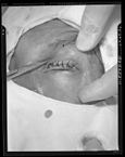
Entropion and trichiasis - By Otis Historical Archives Nat'l Museum of Health & Medicine - originally posted to Flickr as A44-652-11, CC BY 2.0, https://commons.wikimedia.org/w/index.php?curid=6215100

References
- ↑ 1.0 1.1 Dougherty JM, McCulley JP (1986). "Bacterial lipases and chronic blepharitis". Invest Ophthalmol Vis Sci. 27 (4): 486–91. PMID 3957566.
- ↑ 2.0 2.1 2.2 THYGESON P, VAUGHAN DG (1954). "Seborrheic blepharitis". Trans Am Ophthalmol Soc. 52: 173–88. PMC 1312591. PMID 13274422.
- ↑ Lemp MA, Nichols KK (2009). "Blepharitis in the United States 2009: a survey-based perspective on prevalence and treatment". Ocul Surf. 7 (2 Suppl): S1–S14. PMID 19383269.
- ↑ Blepharitis. American Academy of Ophthalmology/eyewiki (2014). http://eyewiki.org/Blepharitis Accessed on July 14, 2016
- ↑ Blepharitis. National Eye Institute. (2009). https://nei.nih.gov/health/blepharitis/blepharitis Accessed on July, 14, 2016
- ↑ Tomlinson A, Bron AJ, Korb DR, Amano S, Paugh JR, Pearce EI; et al. (2011). "The international workshop on meibomian gland dysfunction: report of the diagnosis subcommittee". Invest Ophthalmol Vis Sci. 52 (4): 2006–49. doi:10.1167/iovs.10-6997f. PMC 3072162. PMID 21450918.