Basal lamina
Editor-In-Chief: C. Michael Gibson, M.S., M.D. [1]

The basal lamina is a layer on which epithelium sits and which is secreted by the epithelial cells. It is often confused with the basement membrane, and sometimes used inconsistently in the literature, see below.
It is typically about 40-50 nanometres thick (with exceptions such as the basal laminae that compose the 100-200 nanometre thick glomerular basement membrane).
Layers
The layers of the basal lamina ("BL") and those of the basement membrane ("BM") are described below:
| Name | Part of BL? | Part of BM? | Notes |
| lamina lucida / lamina rara interna[1] | yes | yes | electron-lucid layer[2] containing the glycoprotein laminin |
| lamina densa | yes | yes | electron-dense layer[3] composed of type IV collagen |
| lamina lucida / lamina rara externa | yes | yes | Similar composition to lamina rara interna. Some sources do not consider this a distinct layer. |
| lamina reticularis[4] | no | yes | The three above layers of the basal lamina typically sit on top of the reticular lamina, which is synthesized by cells from the underlying connective tissue and contains fibronectin. The exception is when two epithelial layers abut one another as in the alveoli of the lungs and glomeruli of the kidneys, in which the basal lamina of one epithelial layer fuses with that of the other. |
Anchoring fibers composed of type IV collagen extend from the basal lamina into the underlying reticular lamina and loop around collagen bundles. Although found beneath all basal laminae, they are especially numerous in stratified squamous cells of the skin.
These layers should not be confused with the lamina propria, which is found outside the basal lamina.[5]
Basal lamina vs. basement membrane
The term "basal lamina" is usually used with electron microscopy, while the term "basement membrane" is usually used with light microscopy. The structure known as the basement membrane in light microscopy refers to the stained structure anchoring an epithelial layer. This encompasses the basal lamina secreted by epithelial cells and typically a reticular lamina secreted by other cells.
The basal lamina cannot be distinguished under the light microscope, but under the higher magnification of an electron microscope, the basal lamina and lamina reticularis are visibly distinct structures.
Some theorize that the lamina lucida is an artifact created when preparing the tissue, and that the basement membrane is therefore equal to the lamina densa in vivo.[6]
Examples of basement membranes include:
Additional images
-
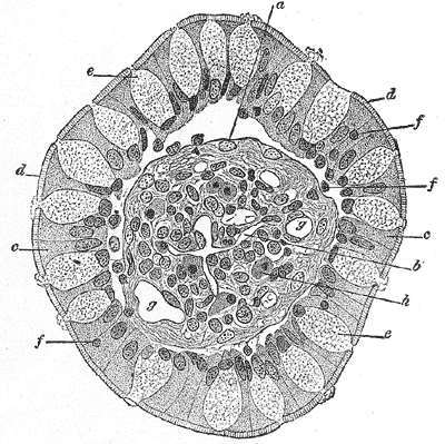
Transverse section of a villus, from the human intestine. X 350.
-

The basal lamina is a component of the basement membrane that separates epithelium from the underlying connective tissue.


See also
References
- ↑ Histology image: 22403loa – Histology Learning System at Boston University
- ↑ Template:UIUCHistologySubject
- ↑ Template:UIUCHistologySubject
- ↑ Histology image: 20904loa – Histology Learning System at Boston University
- ↑ Histology image: 22203loa – Histology Learning System at Boston University
- ↑ Chan F, Inoue S (1994). "Lamina lucida of basement membrane: an artefact". Microsc Res Tech. 28 (1): 48–59. PMID 8061357.
- 1 Kierszenbaum, AL. Histology and Cell Biology: An Introduction to Pathology. Mosby, Inc, MO: 2002. Chapter 4.
External links
- Template:Dorlands - "basal lamina"
- Template:Dorlands - "basement membrane"
- Template:EMedicineDictionary
- Template:AnatomyAtlasesMicroscopic - "Basement Membrane"
- Basement+membrane at the US National Library of Medicine Medical Subject Headings (MeSH)