Atrial septal defect chest x ray: Difference between revisions
No edit summary |
|||
| Line 3: | Line 3: | ||
==Overview== | ==Overview== | ||
Chest x rays may be used as a diagnostic tool in the evaluation of an atrial septal defect. As a modality, chest x rays can be limited in diagnostic imaging quality and may often be elected to be used in conjunction with other imaging techniques. Typical findings may include [[cardiomegaly]] and pulmonary artery enlargement. | |||
== | ==Indications== | ||
* Effectively evaluates the size of the heart | |||
* Effectively evaluates the size of blood vessels involved with pulmonary hemodynamics | |||
Findings of | ==Common Findings== | ||
* In an anteroposterior view, one may see: | |||
:* Prominent pulmonary vascular markings on the chest | |||
:* Enlargement of the cardiac silhouette | |||
:* Normal apperance of the left atrium and left ventricle | |||
:* Triangular appearance of the heart | |||
::* Results from enlargement of pulmonary arteries preventing the ascending and transverse aorta from forming normal heart borders | |||
:* Visible fullness of the right atrium, likely the result of a moderate to large defect | |||
:* [[Scimitar sign]], a vertical, gently curved, right-sided paracardiac density, may be visible | |||
::* Commonly associated with the '''sinus venosus''' atrial septal defect | |||
::* Results from the point of insertion of the pulmonary vein into the inferior vena cava | |||
::* May cause abnormal densities within the chest x ray | |||
* In a lateral view, one may see: | |||
:* Prominent pulmonary vasculature | |||
::*Extends to the periphery of the lung fields and pulmonary trunk | |||
::*Central branches may be dilated | |||
::*Commonly found in '''ostium secundum''' atrial septal defects | |||
* Both views may show [[pulmonary hyperemia]] | |||
==Less Common Findings== | |||
* Normal appereance of heart vasculature | |||
* | * Left heart enlargement/left atrial enlargement | ||
* [[Pulmonary edema]] | |||
* | * Pulmonary venous hypertension | ||
* | |||
==Disadvantages== | |||
* Image quality is not comparable to that of other modalities such as echocardiography and ultrasound | |||
* Appearance of defect signs may be subtle or absent in pediatrics and young adults | |||
====Imaging==== | ====Imaging==== | ||
| Line 34: | Line 54: | ||
[[Category:Cardiology]] | [[Category:Cardiology]] | ||
[[Category:Congenital heart disease]] | [[Category:Congenital heart disease]] | ||
[[Category:Pediatrics]] | |||
[[Category:Mature chapter]] | [[Category:Mature chapter]] | ||
{{WH}} | {{WH}} | ||
{{WS}} | {{WS}} | ||
Revision as of 18:43, 24 August 2011
|
Atrial Septal Defect Microchapters | |
|
Treatment | |
|---|---|
|
Surgery | |
|
| |
|
Special Scenarios | |
|
Case Studies | |
|
Atrial septal defect chest x ray On the Web | |
|
American Roentgen Ray Society Images of Atrial septal defect chest x ray | |
|
Risk calculators and risk factors for Atrial septal defect chest x ray | |
Editor-In-Chief: C. Michael Gibson, M.S., M.D. [1]; Associate Editor(s)-In-Chief: Priyamvada Singh, M.B.B.S. [[2]]; Cafer Zorkun, M.D., Ph.D. [3]; Assistant Editor(s)-In-Chief: Kristin Feeney, B.S. [[4]]
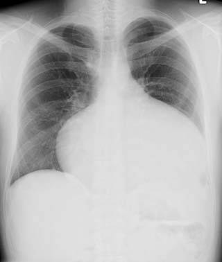
Overview
Chest x rays may be used as a diagnostic tool in the evaluation of an atrial septal defect. As a modality, chest x rays can be limited in diagnostic imaging quality and may often be elected to be used in conjunction with other imaging techniques. Typical findings may include cardiomegaly and pulmonary artery enlargement.
Indications
- Effectively evaluates the size of the heart
- Effectively evaluates the size of blood vessels involved with pulmonary hemodynamics
Common Findings
- In an anteroposterior view, one may see:
- Prominent pulmonary vascular markings on the chest
- Enlargement of the cardiac silhouette
- Normal apperance of the left atrium and left ventricle
- Triangular appearance of the heart
- Results from enlargement of pulmonary arteries preventing the ascending and transverse aorta from forming normal heart borders
- Visible fullness of the right atrium, likely the result of a moderate to large defect
- Scimitar sign, a vertical, gently curved, right-sided paracardiac density, may be visible
- Commonly associated with the sinus venosus atrial septal defect
- Results from the point of insertion of the pulmonary vein into the inferior vena cava
- May cause abnormal densities within the chest x ray
- In a lateral view, one may see:
- Prominent pulmonary vasculature
- Extends to the periphery of the lung fields and pulmonary trunk
- Central branches may be dilated
- Commonly found in ostium secundum atrial septal defects
- Both views may show pulmonary hyperemia
Less Common Findings
- Normal appereance of heart vasculature
- Left heart enlargement/left atrial enlargement
- Pulmonary edema
- Pulmonary venous hypertension
Disadvantages
- Image quality is not comparable to that of other modalities such as echocardiography and ultrasound
- Appearance of defect signs may be subtle or absent in pediatrics and young adults
Imaging
-
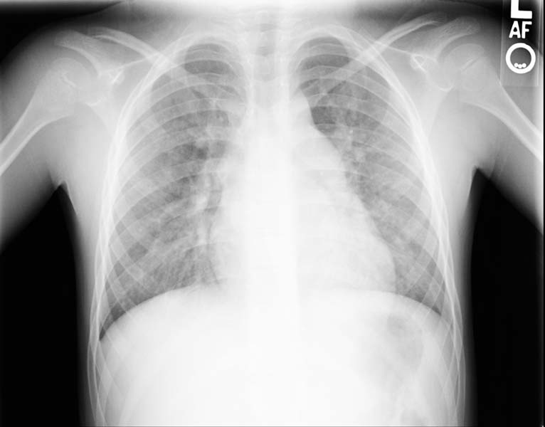
Enlarged right atrial border and mild cardiomegaly.
-
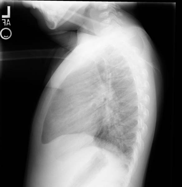
Lateral view
-
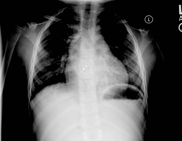
Post repair. Enlarged right atrial border and mild cardiomegaly.
-
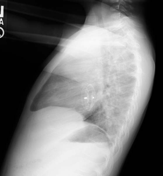
Post repair. Lateral view.
-
ASD. Another patient. Enlarged right atrial border and advanced cardiomegaly.


