Astrocytoma MRI
|
Astrocytoma Microchapters |
|
Diagnosis |
|---|
|
Treatment |
|
Case Study |
|
Astrocytoma MRI On the Web |
|
American Roentgen Ray Society Images of Astrocytoma MRI |
Editor-In-Chief: C. Michael Gibson, M.S., M.D. [1]; Associate Editor(s)-in-Chief: Fahimeh Shojaei, M.D.
Overview
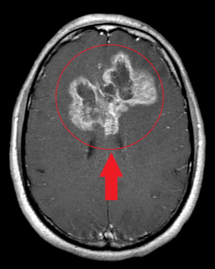
Findings on MRI suggestive of astrocytoma in Low grade astrocytoma (pilocytic and diffuse astrocytoma) include Decreased resonance in comparison to surrounding brain tissue in T1 and Increased resonance in comparison to surrounding brain tissue in T2. In anaplastic astrocytoma we have Hypointense T1, Hyperintense T2 and some contrast enhancement and edema. In glioblastoma multiform we have irregular ring-nodular enhancing lesions and central necrosis surrounding vasogenic edema.
MRI
MRI may be helpful in the diagnosis of astrocytoma. Findings on MRI suggestive of astrocytoma include:[1][2]
- Low grade astrocytoma (pilocytic and diffuse astrocytoma)
- T1: Decreased resonance in comparison to surrounding brain tissue
- T2: Increased resonance in comparison to surrounding brain tissue
- High grade astrocytoma
- Anaplastic astrocytomas
- Hypointense T1
- Hyperintense T2
- There might be some contrast enhancement and edema
- Glioblastoma multiform
- Irregular ring-nodular enhancing lesions
- Central necrosis
- Surrounding vasogenic edema
- Anaplastic astrocytomas
Pilocytic astrocytoma

Diffuse astrocytoma

Anaplastic astrocytoma

Glioblastoma multiform

References
- ↑ Sathornsumetee S, Rich JN, Reardon DA (November 2007). "Diagnosis and treatment of high-grade astrocytoma". Neurol Clin. 25 (4): 1111–39, x. doi:10.1016/j.ncl.2007.07.004. PMID 17964028.
- ↑ Pedersen CL, Romner B (January 2013). "Current treatment of low grade astrocytoma: a review". Clin Neurol Neurosurg. 115 (1): 1–8. doi:10.1016/j.clineuro.2012.07.002. PMID 22819718.
- ↑ Case courtesy of A.Prof Frank Gaillard, <a href="https://radiopaedia.org/">Radiopaedia.org</a>. From the case <a href="https://radiopaedia.org/cases/8474">rID: 8474</a>
- ↑ Case courtesy of Dr Bruno Di Muzio, <a href="https://radiopaedia.org/">Radiopaedia.org</a>. From the case <a href="https://radiopaedia.org/cases/41396">rID: 41396</a>
- ↑ Case courtesy of Dr Bruno Di Muzio, <a href="https://radiopaedia.org/">Radiopaedia.org</a>. From the case <a href="https://radiopaedia.org/cases/39124">rID: 39124</a>
- ↑ Case courtesy of A.Prof Frank Gaillard, <a href="https://radiopaedia.org/">Radiopaedia.org</a>. From the case <a href="https://radiopaedia.org/cases/2589">rID: 2589</a>