|
|
| Line 38: |
Line 38: |
| ==Diagnosis== | | ==Diagnosis== |
|
| |
|
| ===Imaging Findings===
| | [[Arnold-Chiari malformation history and symptoms|History and Symptoms]] | [[Arnold-Chiari malformation physical examination|Physical Examination]] | [[Arnold-Chiari malformation laboratory findings|Laboratory Findings]] | [[Arnold-Chiari malformation x ray|X Ray]] | [[Arnold-Chiari malformation CT|CT]] | [[Arnold-Chiari malformation MRI|MRI]] | [[Arnold-Chiari malformation echocardiography or ultrasound|Echocardiography or Ultrasound]] | [[Arnold-Chiari malformation other imaging findings|Other Imaging Findings]] | [[Arnold-Chiari malformation other diagnostic studies|Other Diagnostic Studies]] |
| ====X ray skull====
| |
| May demonstrate associated abnormalities of the skull base.
| |
| ===CT===
| |
| May demonstrate hydrocephalus, herniated cerebellar tonsils, and a flattened spinal cord. Rarely will CT show a syrinx.
| |
| ===MRI===
| |
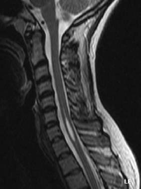
| * [[MRI]] is the imaging modality of choice to evaluate for a Chiari I malformation. MRI findings:
| |
| **[[Cerebellar]] tonsillar herniation
| |
| **Wedge shaped tonsils
| |
| **[[Syringohydromyelia]]
| |
| **Small [[posterior fossa]]
| |
| **Obstructive [[hydrocephalus]]
| |
| **[[Brainstem]] anomalies
| |
| *Tonsillar displacement is measured from the basion-opisthion line on a sigittal image.
| |
| *Herniation is usually at least 5mm, though patients with 3-5mm herniation may also have the malformation.
| |
| *MRI CSF flow studies may prove to be helpful and are currently under investigation.
| |
| | |
| ===Case Study#1===
| |
| Images shown below are courtesy of RadsWiki and copylefted
| |
| | |
| <div align="left">
| |
| <gallery heights="125" widths="125">
| |
| Image:Chiari 1 malformation 001.jpg|Sag T2
| |
| Image:Chiari 1 malformation 002.jpg|Sag T1
| |
| Image:Chiari 1 malformation 003.jpg|Sag T1
| |
| </gallery>
| |
| </div>
| |
| ===Case Study#2===
| |
| | |
| Images shown below are courtesy of RadsWiki and copylefted
| |
| | |
| <div align="left">
| |
| <gallery heights="125" widths="125">
| |
| Image:Chiari 1 malformation 101.jpg|Sag T1
| |
| Image:Chiari 1 malformation 102.jpg|Sag T2
| |
| Image:Chiari 1 malformation 103.jpg|Ax T2
| |
| </gallery>
| |
| </div>
| |
| | |
| | |
| <div align="left">
| |
| <gallery heights="125" widths="125">
| |
| Image:Chiari 1 malformation 104.jpg|Ax T2
| |
| Image:Chiari 1 malformation 105.jpg|Ax T2
| |
| </gallery>
| |
| </div>
| |
| ===Other Radiologic Findings===
| |
| | |
| <div align="left">
| |
| <gallery heights="125" widths="125">
| |
| Image:Arnold-Chiari Malformation MRI 0001.jpg|Brain: Arnold Chiari I; T1 (MRI)
| |
| Image:Arnold-Chiari Malformation CT 0001.jpg|Brain CT: Arnold Chiari I with Intrathecal Contrast; WC
| |
| Image:Arnold-Chiari Malformation MRI 0003.jpg|Brain: Arnold Chiari I, with Hydromyelia of Cervical Spinal Cord; T1 (MRI)
| |
| </gallery>
| |
| </div>
| |
| | |
| | |
| <div align="left">
| |
| <gallery heights="125" widths="125">
| |
| Image:Arnold-Chiari Malformation Plain Film 0001.jpg|Skull: Arnold Chiari II, Skull with Luckenschadel (Plain Film)
| |
| Image:Arnold-Chiari Malformation CT 0002.jpg|Brain: Arnold Chiari II, Hydrocephalus (CT)
| |
| Image:Arnold-Chiari Malformation CT 0003.jpg|Brain: Arnold Chiari II, 6 months Post-Shunt (CT)
| |
| </gallery>
| |
| </div>
| |
|
| |
|
| | ==Treatment== |
|
| |
|
| <div align="left">
| | [[Arnold-Chiari malformation medical therapy|Medical Therapy]] | [[Arnold-Chiari malformation surgery|Surgery]] | [[Arnold-Chiari malformation primary prevention|Primary Prevention]] | [[Arnold-Chiari malformation cost-effectiveness of therapy|Cost-Effectiveness of Therapy]] | [[Arnold-Chiari malformation future or investigational therapies|Future or Investigational Therapies]] |
| <gallery heights="125" widths="125">
| |
| Image:Arnold-Chiari Malformation MRI 0007.jpg|Brain: Arnold Chiari II, with Towering of The Cerebellum and Wide Tentorial Incisure; T1 (MRI)
| |
| Image:Arnold-Chiari Malformation MRI 0008.jpg|Brain: Arnold Chiari II, with Fenestrated Falx Cerebri and Interdigitating Gyri; T2 (MRI)
| |
| Image:Arnold-Chiari Malformation MRI 0009.jpg|Brain: Arnold Chiari II, with Tectal Beaking and Large Massa Intermedia and Stenogyria; T1 (MRI)
| |
| </gallery>
| |
| </div>
| |
| | |
| | |
| <div align="left">
| |
| <gallery heights="125" widths="125">
| |
| Image:Arnold-Chiari Malformation MRI 0010.jpg|Brain: Arnold Chiari Type II with Medullary Spur and Kink (MRI)
| |
| Image:Arnold-Chiari Malformation MRI 0011.jpg|Brain: Arnold Chiari II, with Hypoplastic Corpus Callosum; T1 (MRI)
| |
| Image:Arnold-Chiari Malformation MRI 0012.jpg|Brain: Arnold Chiari II, with Cerebellar Hemispheres that Creep Anteriorly Around the Brain Stem; T1 (MRI)
| |
| </gallery>
| |
| </div>
| |
| | |
| | |
| <div align="left">
| |
| <gallery heights="125" widths="125">
| |
| Image:Arnold-Chiari Malformation MRI 0013.jpg|BRAIN: ARNOLD CHIARI II (MRI)
| |
| Image:Arnold-Chiari Malformation MRI 0014.jpg|BRAIN: ARNOLD CHIARI II WITH ELONGATED FOURTH VENTRICLES AND STENOGYRIA; T1 (MRI)
| |
| Image:Arnold-Chiari Malformation MRI 0015.jpg|BRAIN: ARNOLD CHIARI II WITH HYPOPLASTIC CORPUS CALLOSUM AND STENOGYRIA; T1 (MRI)
| |
| </gallery>
| |
| </div>
| |
| | |
| | |
| <div align="left">
| |
| <gallery heights="125" widths="125">
| |
| Image:Arnold-Chiari Malformation MRI 0016.jpg|BRAIN: ARNOLD CHIARI II WITH REPAIRED MYELOMENINGOCELE AND TETHERED SPINAL CORD; T1 (MRI)
| |
| Image:Arnold-Chiari Malformation MRI 0017.jpg|BRAIN: ARNOLD CHIARI TYPE IV; T1 (MRI)
| |
| Image:Arnold-Chiari Malformation MRI 0018.jpg|BRAIN: ARNOLD CHIARI TYPE IV; T1 (MRI)
| |
| </gallery>
| |
| </div>
| |
| | |
| | |
| <div align="left">
| |
| <gallery heights="125" widths="125">
| |
| Image:Arnold-Chiari Malformation MRI 0019.jpg|BRAIN: ARNOLD CHIARI MALFORMATION II; 1/2 PCMC (MRI)
| |
| Image:Arnold-Chiari Malformation MRI 0020.jpg|BRAIN: ARNOLD CHIARI MALFORMATION II, HERNIATED CEREBELLAR TONSIL; 1/2 PCMC - ARROW (MRI)
| |
| Image:Arnold-Chiari Malformation MRI 0021.jpg|SPINAL CORD: ARNOLD CHIARI MALFORMATION II; 2/2 PCMC (MRI)
| |
| </gallery>
| |
| </div>
| |
| | |
| | |
| <div align="left">
| |
| <gallery heights="125" widths="125">
| |
| Image:Arnold-Chiari Malformation MRI 0022.jpg|SPINAL CORD: ARNOLD CHIARI MALFORMATION II; 2/2 PCMC - ARROW (MRI)
| |
| Image:Arnold-Chiari Malformation MRI 0023.jpg|BRAIN: ARNOLD CHIARI MALFORMATION; 1/3 T2 (MRI)
| |
| Image:Arnold-Chiari Malformation MRI 0024.jpg|BRAIN: ARNOLD CHIARI MALFORMATION; 1/3 - ARROW T2 (MRI)
| |
| </gallery>
| |
| </div>
| |
| | |
| | |
| <div align="left">
| |
| <gallery heights="125" widths="125">
| |
| Image:Arnold-Chiari Malformation MRI 0025.jpg|BRAIN: ARNOLD CHIARI MALFORMATION; 2/3 T2 (MRI)
| |
| Image:Arnold-Chiari Malformation CT 0026.jpg|SPINAL CORD: ARNOLD CHIARI MALFORMATION; 3/3 (CT)
| |
| Image:Arnold-Chiari Malformation CT 0027.jpg|BRAIN: ARNOLD CHIARI MALFORMATION (CT)
| |
| </gallery>
| |
| </div>
| |
| ==Treatment==
| |
| ===Surgery===
| |
| Once these "onset of symptoms" occurs, the most frequent treatment is decompression surgery, in which a neurosurgeon seeks to open the base of the skull and through various methods unrestrict CSF flow to the spine.
| |
|
| |
|
| This treatment is under observation and review. Decompression is a very taxing surgical procedure and is now, in some circles, disdained in lieu of tethered cord detachment at the base of the spine. Some neurological surgeons find that detethering the spinal cord relieves the compression of the brain against the skull opening (foramen magnum) obviating the need for decompression surgery and associated trauma. It should be noted that the alternative spinal surgery is not without risk.
| | ==Case Studies== |
|
| |
|
| ==References==
| | [[Arnold-Chiari malformation case study one|Case #1]] | [[Arnold-Chiari malformation case study two|Case #2]] |
| {{reflist|2}}
| |
|
| |
|
| {{Congenital malformations and deformations of nervous system}} | | {{Congenital malformations and deformations of nervous system}} |
