|
|
| Line 18: |
Line 18: |
|
| |
|
| '''''Synonyms and keywords:''''' ''Chiari malformation''' or '''ACM''' | | '''''Synonyms and keywords:''''' ''Chiari malformation''' or '''ACM''' |
| ==Overview== | | ==[[Arnold-Chiari malformation overview|Overview]]== |
| '''Arnold-Chiari malformation''' is a congenital anomaly of the [[brain]].
| |
| Arnold-Chiari Malformation II occurs in almost all children born with both [[spina bifida]] and [[hydrocephalus]], but ACM I is typically seen in children and adults without spina bifida. The scale of severity is rated I - IV, with IV being the most severe. <ref>Barkovich AJ, Wippold FJ, Sherman JL, Citrin CM. [http://www.ajnr.org/cgi/content/abstract/7/5/795 Significance of cerebellar tonsillar position on MR.] AJNR Am J Neuroradiol. 1986 Sep-Oct;7(5):795-9.</ref> <ref>Haughton VM, Korosec FR, Medow JE, Dolar MT, Iskandar BJ. [http://www.ajnr.org/cgi/content/abstract/24/2/169 Peak systolic and diastolic CSF velocity in the foramen magnum in adult patients with Chiari I malformations and in normal control participants.] AJNR Am J Neuroradiol. 2003 Feb;24(2):169-76.</ref> <ref>Elster A, Chen M. [http://radiology.rsnajnls.org/cgi/content/abstract/183/2/347 Chiari I malformations: clinical and radiologic reappraisal.] Radiology. 1992; 183:347-353.</ref> | |
| ==Historical Perspective==
| |
| An Austrian [[pathologist]], [[Hans Chiari]], first described these hindbrain malformations in the 1890s. A colleague of Professor Chiari, Dr. Julius Arnold later contributed to the definition of the condition, and students of Dr. Arnold suggested the term "Arnold-Chiari malformation" to henceforth refer to the condition.<ref>{{WhoNamedIt|synd|1154|Arnold-Chiari malformation}}</ref>
| |
| ==Classification Scheme== | |
| The Austrian pathologist Hans Chiari in the late 19th century described seemingly related anomalies of the hindbrain, the so called Chiari malformations I, II and III. Later, other investigators added a fourth (Chiari IV) malformation. The scale of severity is rated I - IV, with IV being the most severe. Types III and IV are very rare.<ref name="urlArnold Chiari Malformation">{{cite web|url=http://neurosurgery.ucla.edu/body.cfm?id=109 |title=Arnold Chiari Malformation |work= |accessdate=}}</ref>
| |
|
| |
|
| {| class="wikitable"
| | ==[[Arnold-Chiari malformation historical perspective|Historical Perspective]]== |
| |-
| |
| ! Type
| |
| ! Presentation
| |
| ! Other notes
| |
| |-
| |
| | I
| |
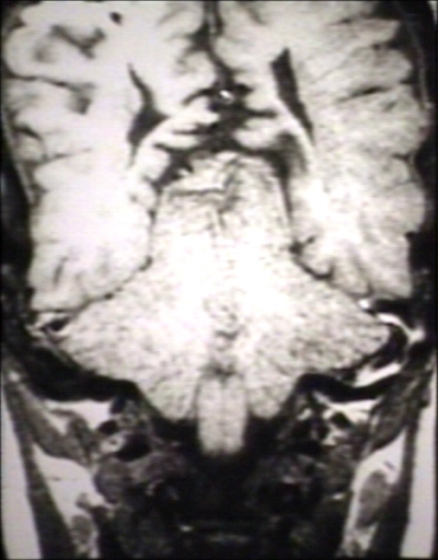
| | A congenital malformation. Is generally asymptomatic during childhood, but often manifests with headaches and cerebellar symptoms. Herniation of [[cerebellar tonsil]]s.<ref name="pmid19246872">{{Cite journal|author=Kojima A, Mayanagi K, Okui S |title=Progression of pre-existing Chiari type I malformation secondary to cerebellar hemorrhage: case report |journal=Neurol. Med. Chir. (Tokyo) |volume=49 |issue=2 |pages=90–2 |year=2009 |month=February |pmid=19246872 |doi= 10.2176/nmc.49.90|url=http://joi.jlc.jst.go.jp/JST.JSTAGE/nmc/49.90?from=PubMed |format=}} {{Dead link|date=May 2009}}</ref><ref name="pmid16509477">{{Cite journal|author=O'Shaughnessy BA, Bendok BR, Parkinson RJ, ''et al.'' |title=Acquired Chiari malformation Type I associated with a supratentorial arteriovenous malformation. Case report and review of the literature |journal=J. Neurosurg. |volume=104 |issue=1 Suppl |pages=28–32 |year=2006 |month=January |pmid=16509477 |doi=10.3171/ped.2006.104.1.28 |url=http://thejns.org/doi/abs/10.3171/ped.2006.104.1.28?url_ver=Z39.88-2003&rfr_id=ori:rid:crossref.org&rfr_dat=cr_pub%3dncbi.nlm.nih.gov}}</ref>
| |
| | The most common form.
| |
| |-
| |
| |Syndrome of occipitoatlantoaxial hypermobility
| |
| |An acquired Chiari I Malformation in patients with hereditary disorders of connective tissue.<ref name="Milhorat-2007"/> Patients who exhibit extreme joint [[hypermobility]] and connective tissue weakness as a result of [[Ehlers-Danlos syndrome]] or [[Marfan Syndrome]] are susceptible to instabilities of the craniocervical junction and thus acquiring a Chiari Malformation. This type is difficult to diagnose and treat.<ref name="Conquer Chiari, Connective Tissue Disorders">{{cite web|url=http://www.conquerchiari.org/subs%20only/volume%204/issue%204(10)/bland%20connective%20tissue%204(10).asp |title=Dr. Bland Discusses Chiari & EDS 4(10) |publisher=Conquerchiari.org |date=2006-11-20 |accessdate=2011-11-04}}</ref>
| |
| |
| |
| |-
| |
| | II
| |
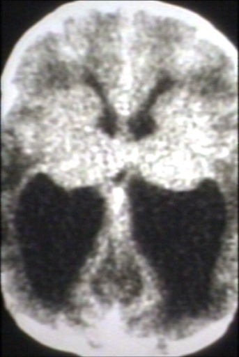
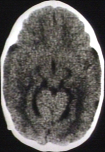
| | Usually accompanied by a [[lumbar]] [[myelomeningocele]]<ref name="urlNeuroradiology - Chiari malformation (I-IV)">{{cite web|url=http://www.mir.wustl.edu/neurorad/internal.asp?NavID=123 |title=Neuroradiology - Chiari malformation (I-IV) |work= |accessdate=}}</ref> leading to partial or complete paralysis below the spinal defect. As opposed to the less pronounced tonsillar herniation seen with Chiari I, there is a larger cerebellar vermian displacement. Low lying [[torcular herophili]], tectal beaking, and hydrocephalus with consequent clival hypoplasia are classic anatomic associations.<ref>{{cite web|url =http://www.cchs.net/pediatricradiology/imagegallery/default.asp|title =Pediatric Radiology Image Gallery|accessdate = June 14, 2010|year = 2010|title = Cleveland Clinic Children's Hospital Pediatric Radiology Image Gallery|accessdate = June 14, 2010|publisher = [[Cleveland Clinic]]| archiveurl= http://web.archive.org/web/20100627053310/http://www.cchs.net/pediatricradiology/imagegallery/default.asp| archivedate= 27 June 2010 <!--DASHBot-->| deadurl= no}}</ref> The position of the torcular herophili is important for distinction from [[Dandy-Walker syndrome]] in which it is classically upturned. This is important because the hypoplastic cerebellum of Dandy-Walker may be difficult to distinguish from a Chiari malformation that has herniated or is ectopic on imaging. [[Colpocephaly]] may be seen due to the associated neural tube defect.
| |
| |
| |
| |-
| |
| | III
| |
| | Causes severe neurological defects. It is associated with an [[occiput|occipital]] [[encephalocele]].<ref name="Mesh">{{MeshName|Arnold-Chiari+Malformation}}</ref>
| |
| |
| |
| |-
| |
| | IV
| |
| | Characterized by a lack of [[cerebellar]] development.<ref name="urlChiari Malformations - Department of Neurological Surgery">{{cite web|url=http://www.cumc.columbia.edu/dept/nsg/ct/chiari_malformation.html |title=Chiari Malformations - Department of Neurological Surgery |work= |accessdate=}}</ref>
| |
| |
| |
| |}
| |
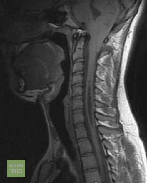
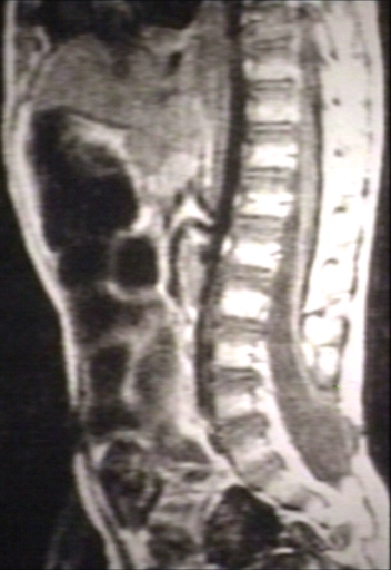
| [[File:Neck MRI 130850-dichromatic t1-t2-t2.png|thumb|200px|Syringomiyelia associated with Chiari malformation]]
| |
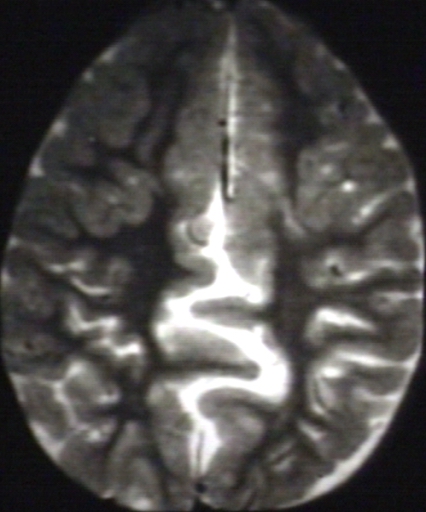
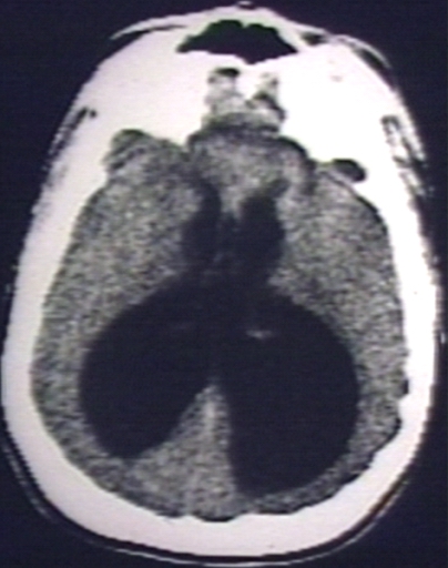
| Other conditions sometimes associated with Chiari Malformation include [[hydrocephalus]],<ref name="urlNeuropathology For Medical Students">{{cite web|url=http://www.pathology.vcu.edu/WirSelfInst/neuro_medStudents/devdis.html |title=Neuropathology For Medical Students |work= |accessdate=}}</ref> [[syringomyelia]], [[spinal curvature]], [[tethered spinal cord syndrome]], and connective tissue disorders<ref name="Milhorat-2007">{{Cite journal|author=Milhorat TH, Bolognese PA, Nishikawa M, McDonnell NB, Francomano CA |title=Syndrome of occipitoatlantoaxial hypermobility, cranial settling, and chiari malformation type I in patients with hereditary disorders of connective tissue |journal=[[Journal of Neurosurgery|Journal of Neurosurgery: Spine]] |volume=7 |issue=6 |pages=601–9 |year=2007 |month=December |pmid=18074684 |doi=10.3171/SPI-07/12/601 |url=http://thejns.org/doi/full/10.3171/SPI-07/12/601}}</ref> such as [[Ehlers-Danlos syndrome]] and [[Marfan Syndrome]].
| |
| ==Pathophysiology==
| |
| *The most widely accepted pathophysiological mechanism by which Chiari Type 1 Malformations occur is by a reduction or lack of development of the [[posterior fossa]] as a result of either congenital or acquired disorders.
| |
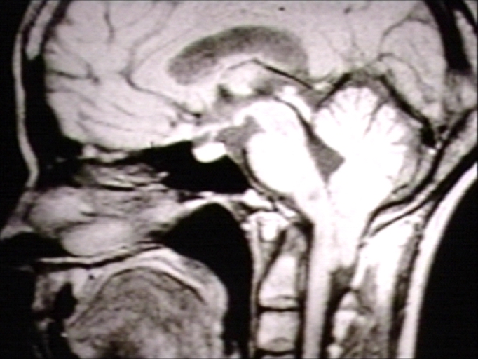
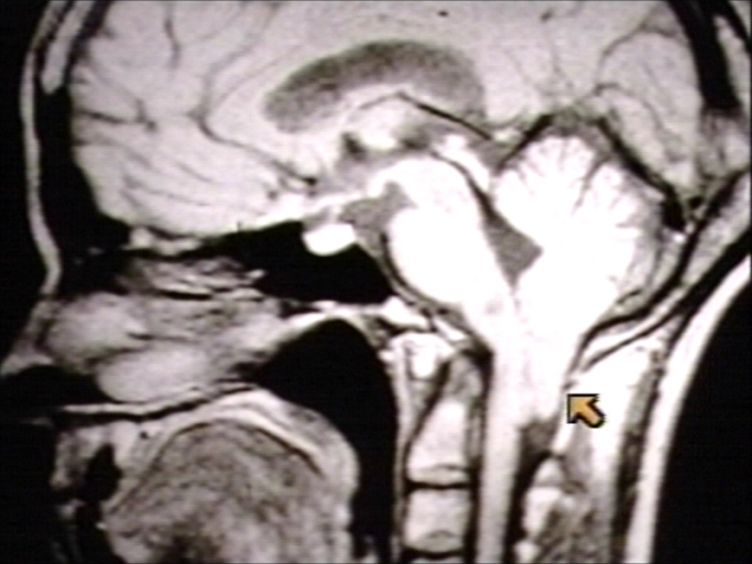
| *The [[cerebellar tonsils]] are elongated and pushed down through the opening of the base of the [[skull]] (see [[foramen magnum]]), blocking the flow of [[cerebrospinal fluid]] (CSF).
| |
| *The [[brainstem]], cranial nerves, and the lower portion of the [[cerebellum]] may be stretched or compressed.
| |
| *Therefore, any of the functions controlled by these areas may be affected. The blockage of CSF flow may also cause a [[syrinx (medicine)|syrinx]] to form, eventually leading to [[syringomyelia]]. Many sufferers turn to the Chiari Institute in Long Island, NY for specialized medical attention and medication.
| |
| ===Gross Pathology===
| |
|
| |
|
| <div align="left">
| | ==[[Arnold-Chiari malformation classification|Classification]]== |
| <gallery heights="125" widths="125">
| |
| Image:Arnold-Chiari Malformation 0001.jpg|Brain: Arnold-Chiari Malformation: Gross fixed tissue sagittal section brain stem
| |
| Image:Arnold-Chiari Malformation 0002.jpg|Brain: Arnold Chiari Malformation: Gross fixed tissue sagittal section cerebrum brainstem and cerebellum
| |
| Image:Arnold-Chiari Malformation 0003.jpg|Brain: Arnold Chiari Malformation And Polygyria: Gross fix tissue external view
| |
| </gallery>
| |
| </div>
| |
|
| |
|
| | ==[[Arnold-Chiari malformation pathophysiology|Pathophysiology]]== |
|
| |
|
| <div align="left">
| | ==[[Arnold-Chiari malformation causes|Causes]]== |
| <gallery heights="125" widths="125">
| |
| Image:Arnold-Chiari Malformation 0004.jpg|Brain: Arnold Chiari Malformation: Gross fixed tissue sagittal section brainstem close-up
| |
| Image:Arnold-Chiari Malformation 0005.jpg|Brain: Arnold Chiari Malformation: Gross fixed tissue brain stem sagittal section close-up
| |
| Image:Arnold-Chiari Malformation 0006.jpg|Brain: Arnold Chiari Malformation: Gross fixed tissue sagittal section brain stem and cerebellum
| |
| </gallery>
| |
| </div>
| |
|
| |
|
| | ==[[Arnold-Chiari malformation differential diagnosis|Differentiating Arnold-Chiari malformation from other Diseases]]== |
|
| |
|
| <div align="left">
| | ==[[Arnold-Chiari malformation epidemiology and demographics|Epidemiology and Demographics]]==] |
| <gallery heights="125" widths="125">
| |
| Image:Arnold-Chiari Malformation 0007.jpg|Brain: Arnold Chiari Malformation: Gross fixed tissue sagittal section brain stem cerebellum and spinal column
| |
| Image:Arnold-Chiari Malformation 0008.jpg|Brain: Hydrocephalus Secondary To Arnold Chiari Malformation: Gross fixed tissue three coronal sections cerebral hemispheres
| |
| Image:Arnold-Chiari Malformation 0009.jpg|Brain: Arnold Chiari Malformation: Gross fixed tissue cerebellum and brainstem
| |
| </gallery>
| |
| </div>
| |
|
| |
|
| | ==[[Arnold-Chiari malformation risk factors|Risk Factors]]== |
|
| |
|
| <div align="left">
| | ==[[Arnold-Chiari malformation natural history, complications and prognosis|Natural History, Complications and Prognosis]]== |
| <gallery heights="125" widths="125">
| |
| Image:Arnold-Chiari Malformation 0010.jpg|Spinal cord: Malformation Vertebral Bodies: Gross natural color sagittal section spinal column with malformation in region C7 T1 associated with Arnold Chiari malformation
| |
| Image:Arnold-Chiari Malformation 0011.jpg|Brain: Arnold Chiari Malformation; with Hydrocephalus, Type I
| |
| Image:Arnold-Chiari Malformation 0012.jpg|Brain: Arnold Chiari Malformation; Type II
| |
| </gallery>
| |
| </div>
| |
|
| |
|
|
| |
| <div align="left">
| |
| <gallery heights="125" widths="125">
| |
| Image:Arnold-Chiari Malformation 0013.jpg|Brain: Arnold Chiari Type II with Meningomyelocele
| |
| Image:Arnold-Chiari Malformation 0014.jpg|Brain: Arnold Chiari Malformation; Mid Sagittal
| |
| Image:Arnold-Chiari Malformation 0015.jpg|Brain: Arnold Chiari Malformation with Myelocele
| |
| </gallery>
| |
| </div>
| |
|
| |
|
| |
| <div align="left">
| |
| <gallery heights="125" widths="125">
| |
| Image:Arnold-Chiari Malformation 0016.jpg|Brain: Arnold Chiari Malformation with Hydrocephalus
| |
| Image:Arnold-Chiari Malformation 0017.jpg|Brain: Arnold Chiari Malformation with Hydrocephalus
| |
| Image:Arnold-Chiari Malformation 0018.jpg|Brain: Arnold Chiari Malformation
| |
| </gallery>
| |
| </div>
| |
|
| |
|
| |
| <div align="left">
| |
| <gallery heights="125" widths="125">
| |
| Image:Arnold-Chiari Malformation 0019.jpg|Brain: Arnold Chiari Malformation
| |
| Image:Arnold-Chiari Malformation 0020.jpg|Brain: Arnold Chiari Malformation
| |
| Image:Arnold-Chiari Malformation 0021.jpg|Brain: Arnold Chiari Malformation, a close up view
| |
| </gallery>
| |
| </div>
| |
|
| |
|
| |
| <div align="left">
| |
| <gallery heights="125" widths="125">
| |
| Image:Arnold-Chiari Malformation 0022.jpg|Brain: Arnold Chiari Malformation; Tonsilar Herniation of Cerebellum Compressing Medulla
| |
| Image:Arnold-Chiari Malformation 0023.jpg|Brain: Arnold Chiari Malformation; Meningomyelocele, Type II, Meningitis
| |
| Image:Arnold-Chiari Malformation 0024.jpg|Brain: Arnold Chiari Malformation, Type II; Meningomyelocele, Meningitis, Close-up of Previous one
| |
| </gallery>
| |
| </div>
| |
|
| |
|
| |
| <div align="left">
| |
| <gallery heights="125" widths="125">
| |
| Image:Arnold-Chiari Malformation 0025.jpg|Brain: Arnold Chiari Malformation, Intramedullary Hemorrhage
| |
| Image:Arnold-Chiari Malformation 0026.jpg|Brain: Arnold Chiari Malformation, Type II
| |
| Image:Arnold-Chiari Malformation 0027.jpg|Spinal Cord: Arnold Chiari Malformation; Type II, Meningomyelitis
| |
| </gallery>
| |
| </div>
| |
|
| |
|
| |
| <div align="left">
| |
| <gallery heights="125" widths="125">
| |
| Image:Arnold-Chiari Malformation 0028.jpg|Spinal Cord: Arnold Chiari Malformation; Type II, Meningomyelitis
| |
| Image:Arnold-Chiari Malformation 0029.jpg|Brain: Arnold Chiari Malformation
| |
| Image:Arnold-Chiari Malformation 0030.jpg|Brain: Arnold Chiari Malformation; Note Z-Shaped Kink in Cervical Spinal Cord
| |
| </gallery>
| |
| </div>
| |
|
| |
|
| |
| <div align="left">
| |
| <gallery heights="125" widths="125">
| |
| Image:Arnold-Chiari Malformation 0031.jpg|Brain: Arnold Chiari Malformation with Hydrocephalus
| |
| Image:Arnold-Chiari Malformation 0032.jpg|Skin: Myelomeningocele with Hydrocephalus; Arnold Chiari; Note Underdevelopment of Legs
| |
| </gallery>
| |
| </div>
| |
|
| |
|
| |
| <div align="left">
| |
| <gallery heights="125" widths="125">
| |
| Image:Arnold-Chiari Malformation 0033.jpg|Brain: Cerebellum Arnold Chiari: Gross fixed tissue flattened cerebellum
| |
| Image:Arnold-Chiari Malformation 0034.jpg|Brain: Polymicrogyria: Gross fixed brain lateral view of left cerebral hemisphere case of Arnold Chiari cerebellum
| |
| </gallery>
| |
| </div>
| |
| ==Causes==
| |
| Congenital causes are
| |
| * [[Craniosynostosis]] (especially of the lambdoid suture)
| |
| * [[Hyperostosis]] (ex. craniometaphyseal dysplasia
| |
| * [[Osteopetrosis]]
| |
| * [[X-linked vitamin D-resistant rickets]]
| |
| * [[Neurofibromatosis type I]]
| |
|
| |
| Acquired disorders include space occupying lesions due to one of several potential causes ranging from brain tumors to hematomas.<ref name="pmid21882908">{{Cite journal|author=Loukas M, Shayota BJ, Oelhafen K, Miller JH, Chern JJ, Tubbs RS, Oakes WJ |title=Associated disorders of Chiari Type I malformations: a review |journal=Neurosurg Focus |volume=31 |issue=3 |pages=E3 |year=2011 |pmid=21882908 |doi=10.3171/2011.6.FOCUS11112 |url=http://thejns.org/doi/abs/10.3171/2011.6.FOCUS11112}}</ref>
| |
| ==Diagnosis
| |
| ==Diagnosis== | | ==Diagnosis== |
| ===Symptoms===
| |
| In [[infant]]s, the most common symptoms are
| |
| * [[Stridor]]
| |
| * [[Swallowing]] difficulties
| |
|
| |
| In older children
| |
| * Upper (and lower as age marches on) limb weakness
| |
| * [[Breathing difficulties]] may occur
| |
| * Patients may experience no symptoms or remain asymptomatic until early adulthood at which point they will often experience severe headaches and neck pain
| |
| * [[Fatigue]]
| |
| * [[Dizziness]]
| |
| * [[Vertigo]]
| |
| * [[Neuropathic pain]]
| |
| * [[Pain]] at the point of tethering
| |
| * [[Visual]] disturbances
| |
| * Difficulty [[swallowing]]
| |
| * Ringing in the ears
| |
| * [[Sleep apnea]]
| |
| * Impaired [[fine motor]] skills
| |
| * Muscle weakness
| |
| * [[Palpitations]]
| |
| * Excessive clearing of the throat with no obstructions
| |
| * Because of the complex combination of symptoms and the lack of experience with ACM1 had by many, even outstanding neurologists and neurosurgeons, many patients are frequently misdiagnosed.
| |
| * Some patients may go an entire lifetime without having noticeable symptoms. Or, symptoms can be minimal, then turn severe suddenly due to head trauma which alters the condition of the spine, brain, or cerebellar tonsils and begins to cause more difficulties...
| |
|
| |
|
| ===Physical Examination===
| |
| ===Vitals===
| |
| ====Pulse====
| |
| ====Rate====
| |
| [[Tachycardia]] may be present.
| |
| ===Eye===
| |
| *[[Nystagmus]] (irregular eye movements) may be present.
| |
| *[[Pupillary dilatation]]
| |
| ====Neurologic====
| |
| *Impaired [[gag reflex]]
| |
| *Impaired [[coordination]]
| |
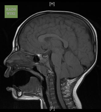
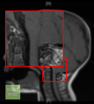
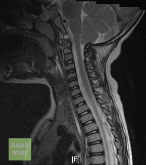
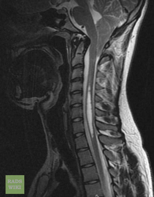
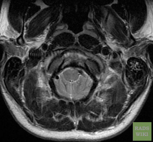
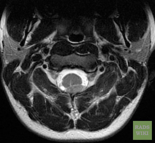
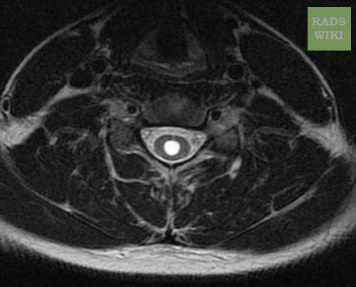
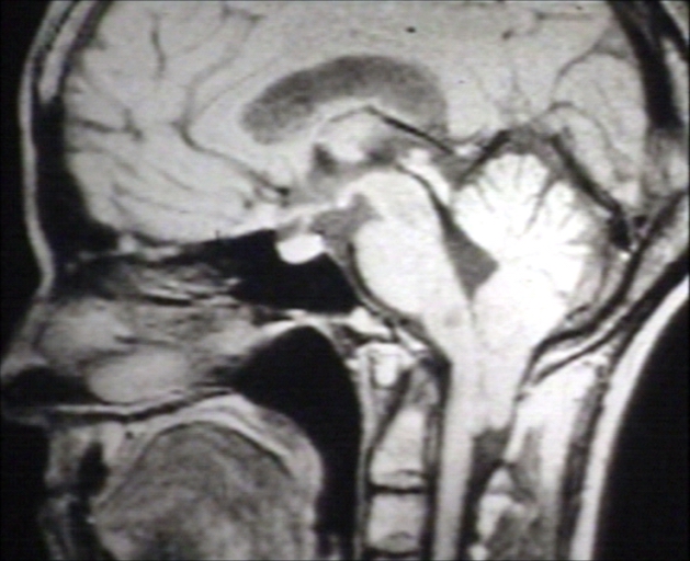
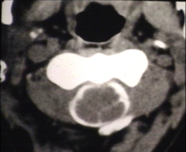
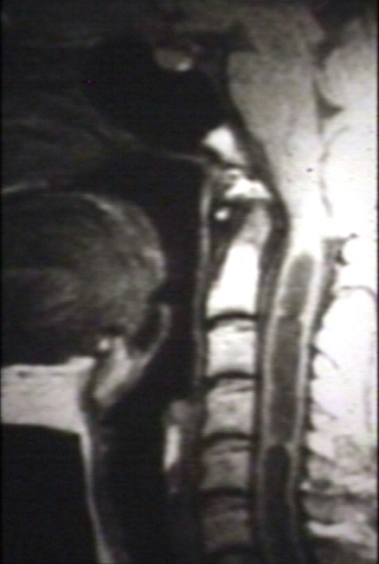
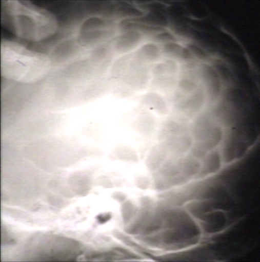
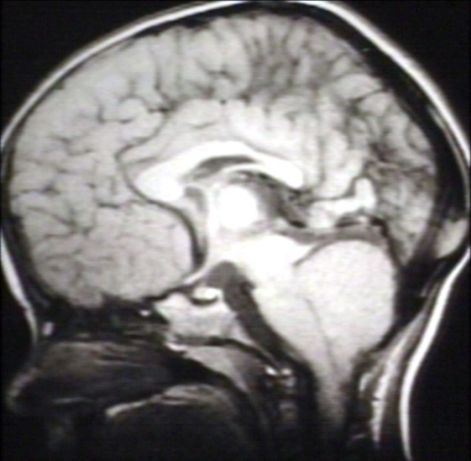
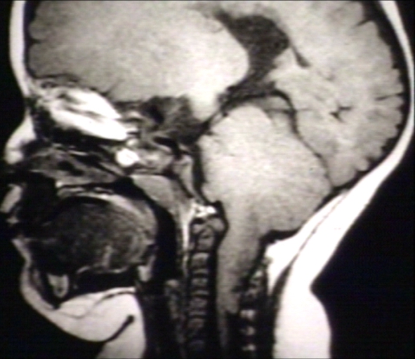
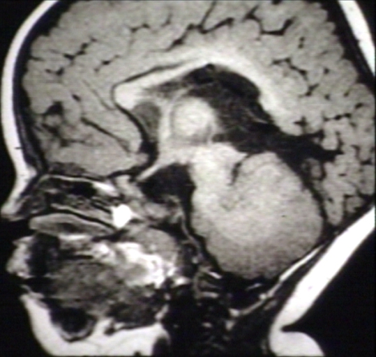
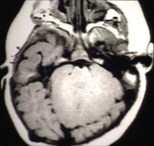
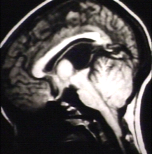
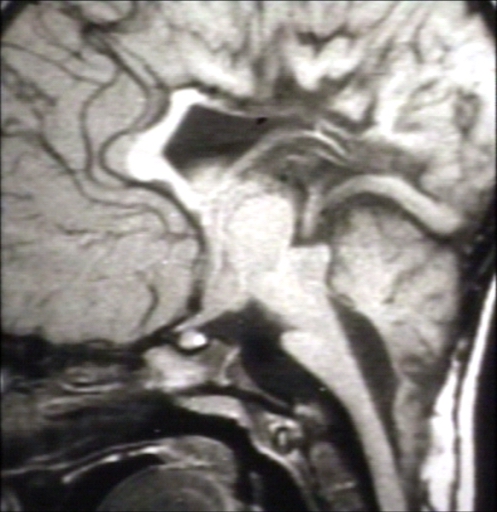
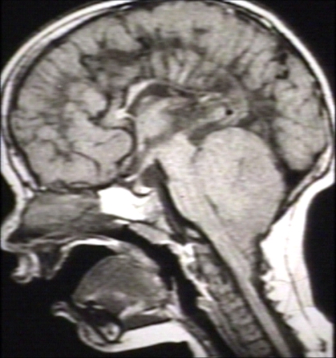
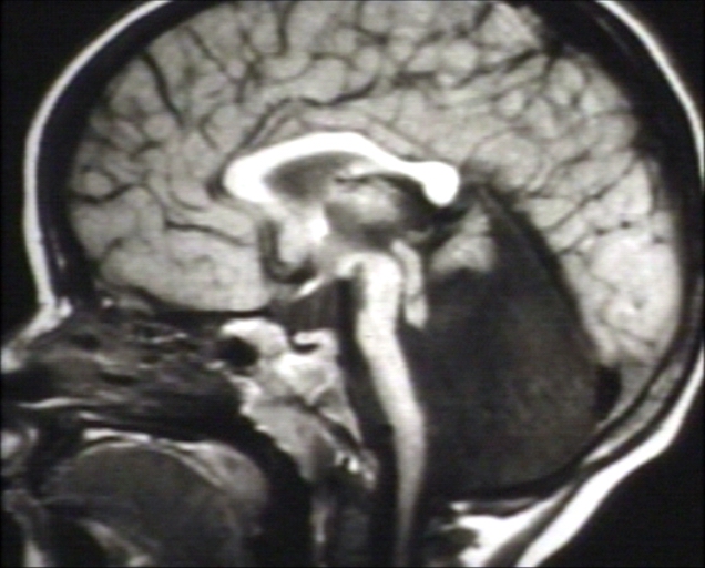
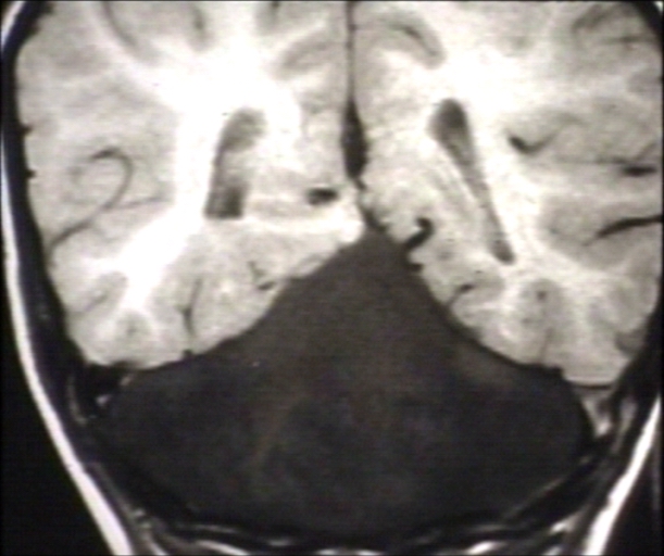
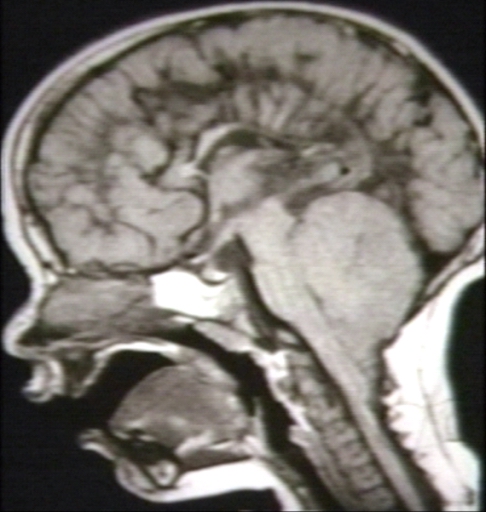
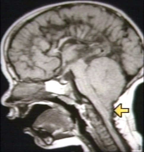
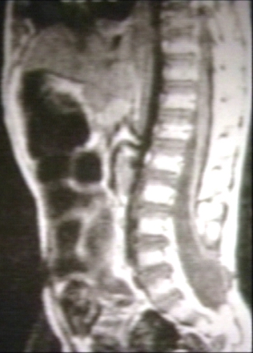
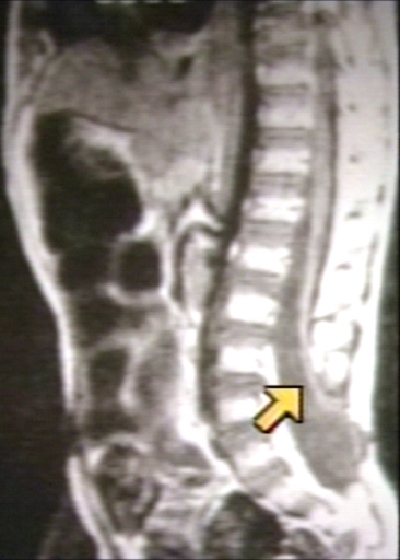
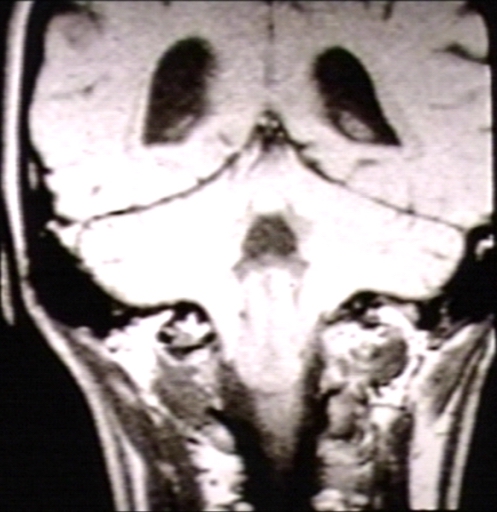
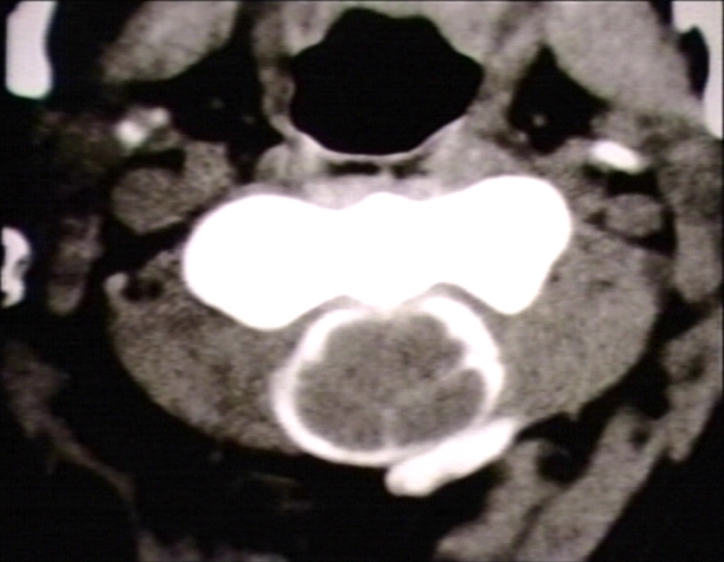
| ===Imaging Findings=== | | ===Imaging Findings=== |
| ====X ray skull==== | | ====X ray skull==== |