Arachnoid cyst MRI: Difference between revisions
Jose Loyola (talk | contribs) No edit summary |
Jose Loyola (talk | contribs) No edit summary |
||
| Line 41: | Line 41: | ||
| | | | ||
|} | |} | ||
* As differential diagnosis, the following hypothesis must be considered: | |||
enlarged CSF space (e.g. mega cisterna magna) | |||
epidermoid cyst | |||
often shows a heterogeneous/dirty signal on FLAIR | |||
restricted diffusion | |||
more lobulated | |||
tend to engulf adjacent arteries and cranial nerves | |||
subdural hygroma/chronic subdural hemorrhage | |||
do not typically show CSF signal intensity on MRI | |||
can have an enhancing membrane | |||
cystic tumors: often will have a solid/enhancing component and be intra-axial | |||
pilocytic astrocytoma | |||
hemangioblastoma | |||
non-neoplastic cysts | |||
neurenteric cyst | |||
neuroglial cyst | |||
porencephalic cyst | |||
often follow a history of trauma or stroke | |||
surrounded by gliotic brain | |||
neurocysticercosis | |||
small cyst | |||
usually multiple when in the subarachnoid space | |||
==References== | ==References== | ||
{{Reflist|2}} | {{Reflist|2}} | ||
==MRI Examples of Arachnoid Cysts | ==MRI Examples of Arachnoid Cysts== | ||
([http://www.radswiki.net Images courtesy of RadsWiki]) | ([http://www.radswiki.net Images courtesy of RadsWiki]) | ||
Revision as of 21:24, 26 June 2020
|
Arachnoid cyst Microchapters |
|
Diagnosis |
|---|
|
Treatment |
|
Case Studies |
|
Arachnoid cyst MRI On the Web |
|
American Roentgen Ray Society Images of Arachnoid cyst MRI |
Editor-In-Chief: C. Michael Gibson, M.S., M.D. [1] Associate Editor(s)-in-Chief: José Eduardo Riceto Loyola Junior, M.D.[2]
Overview
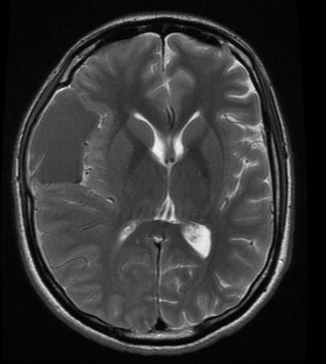
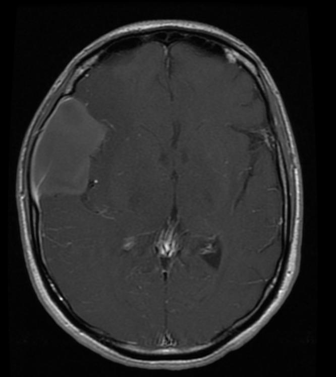
On brain/spine MRI, arachnoid cysts are characterized by cystic images with similar density to CSF and non-enhancing borders, mostly found in the middle cranial fossa while they only rarely occur in the spinal cord. MRIs are more adequate than CT scans for evaluating arachnoid cysts.
MRI
- MRIs are better to diagnose and evaluate the extent of the arachnoid cyst than the CT Scan;
- Most cysts (50-60%) are found in the floor of the middle cranial fossa, while 1/4 to 1/3 of occur in the posterior fossa, particularly in the retrocerebellar, cerebellopontine, and quadrigeminal plate cisterns. Rarely, they may be found in the spinal cord.[1][2]
- Demonstrate the exact location, extent, and relationship of the cyst;
- Can differentiate arachnoid from epidermoid cysts (arachnoid cysts are identical to CSF, while epidermoid present a higher signal with FLAIR and reduced diffusion with DWI, making them appear brighter than CSF).
- CSF signal is seen within the cyst;
- Eventually, arachnoid cysts may contain proteinaceous fluid or blood, which can cause diagnostic confusion.[3]
Differential Diagnosis
| Intraventricularly: | Colloid cysts |
| Intraparenchymally: | Parasitic infections, cystic metastases |
| Porencephalic cysts | |
| Craniopharyngiomas | |
| Holoprosencephalies | |
| Agenesis of corpus callosum | |
| Defect in the hemispheral cleavage | |
| Dandy-Walker complex (posterior fossa cysts) |
- As differential diagnosis, the following hypothesis must be considered:
enlarged CSF space (e.g. mega cisterna magna) epidermoid cyst often shows a heterogeneous/dirty signal on FLAIR restricted diffusion more lobulated tend to engulf adjacent arteries and cranial nerves subdural hygroma/chronic subdural hemorrhage do not typically show CSF signal intensity on MRI can have an enhancing membrane cystic tumors: often will have a solid/enhancing component and be intra-axial pilocytic astrocytoma hemangioblastoma non-neoplastic cysts neurenteric cyst neuroglial cyst porencephalic cyst often follow a history of trauma or stroke surrounded by gliotic brain neurocysticercosis small cyst usually multiple when in the subarachnoid space
References
- ↑ Robertson, S. J., S. M. Wolpert, and V. M. Runge. "MR imaging of middle cranial fossa arachnoid cysts: temporal lobe agenesis syndrome revisited." American journal of neuroradiology 10.5 (1989): 1007-1010.
- ↑ "Arachnoid Cysts - Imaging". Medscape. 06/26/2020. Check date values in:
|date=(help) - ↑ "Arachnoid Cysts". MedPix. 06/26/2020. Check date values in:
|date=(help) - ↑ Cincu, Rafael, Amit Agrawal, and Jose Eiras. "Intracranial arachnoid cysts: current concepts and treatment alternatives." Clinical neurology and neurosurgery 109.10 (2007): 837-843.
MRI Examples of Arachnoid Cysts
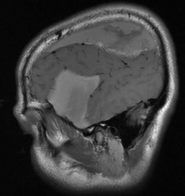
Patient #1: Left middle cranial fossa arachnoid cyst
-
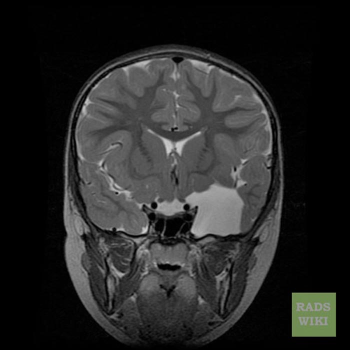
Cor T2
-
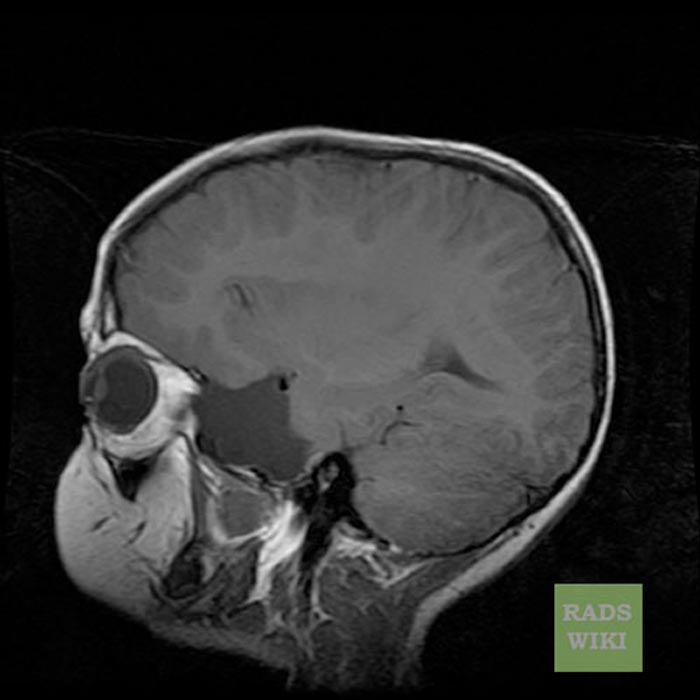
Sag T1
-
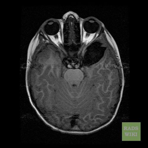
Axial T1 FLAIR
-
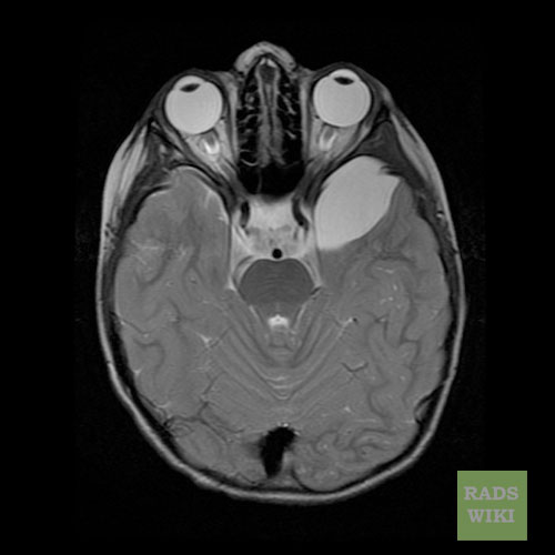
Axial T2


Patient #2: CT and MR images demonstrate a hemorrhagic arachnoid cyst
-
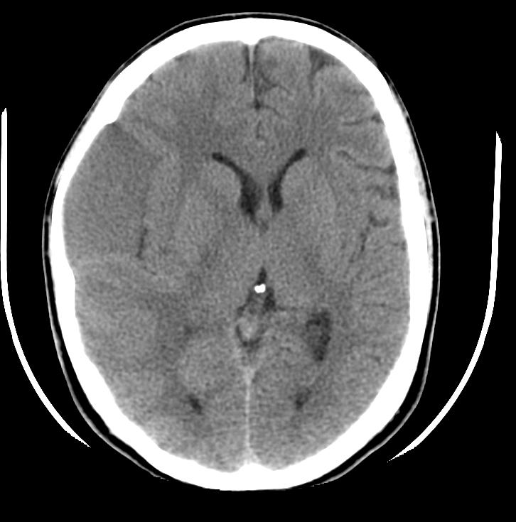
CT
-
Sag T1
-
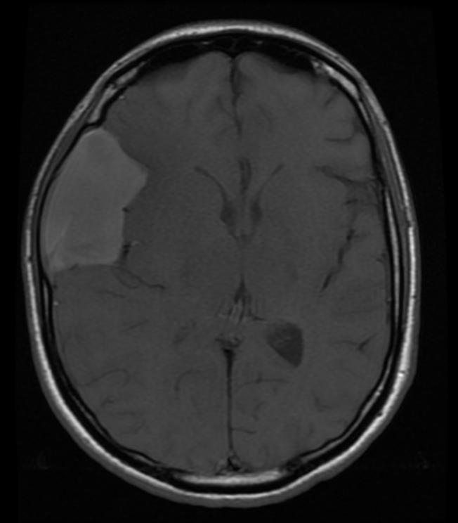
Ax T1
-
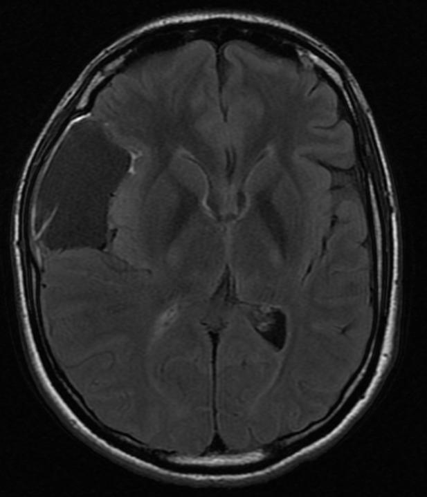
Ax FLAIR
-
Ax T2
-
Ax T1 with GAD


